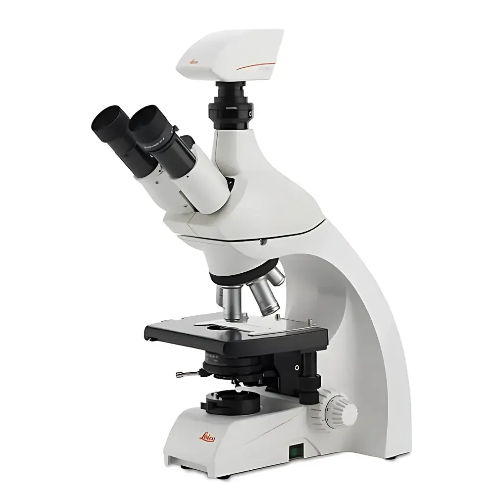
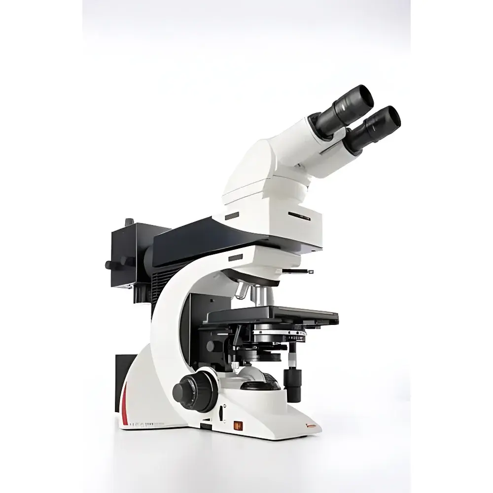
Leica DM1000 Research-Grade Biological Microscope
| Brand | Leica |
|---|---|
| Origin | Germany |
| Model | DM1000 |
| Type | Upright Biological Microscope |
| Optical Design | HC Infinity-Corrected Optics |
| Illumination | 12 V, 30 W Halogen with Köhler Illumination |
| Objective Turret | 5-Position |
| Focus Mechanism | Coaxial coarse/fine focus with 1 µm minimum step resolution |
| Stage | Ceramic-coated mechanical stage with ergonomic controls |
| Eyepieces | Widefield 10×/22 mm, diopter-adjustable |
| Objectives | HC PL Fluotar 4×/0.10, 10×/0.25, 20×/0.40, 40×/0.65, 100×/1.25 Oil (Phase Contrast Capable) |
| Condenser | Adjustable Abbe condenser with color-coded aperture diaphragm |
| Camera Interface | Trinocular port compatible with Leica DFC290 FireWire (IEEE 1394) digital camera (7.15 MP, 3073 × 2304, 3.2 µm pixel size, 30-bit color depth, up to 25 fps at 1024 × 768) |
Overview
The Leica DM1000 is a research-grade upright biological microscope engineered for high-fidelity routine and advanced life science applications—including brightfield, phase contrast, and optional fluorescence imaging. Built upon Leica’s HC (High Contrast) infinity-corrected optical platform, the system delivers consistent image fidelity across magnifications while minimizing chromatic and spherical aberrations. Its Köhler illumination architecture ensures uniform, glare-free specimen illumination—critical for quantitative morphological analysis and long-duration observation sessions. The DM1000 features a thermally compensated mechanical body design that maintains focal plane stability during extended use, reducing drift-induced refocusing and supporting reproducible positional documentation in time-lapse or multi-user lab environments.
Key Features
- HC infinity-corrected optical path with high-transmission coatings optimized for visible-light contrast and resolution
- Five-position objective turret accommodating standard phase contrast objectives: 4×/0.10, 10×/0.25, 20×/0.40, 40×/0.65, and 100×/1.25 oil immersion (all PL Fluotar series)
- Coaxial coarse/fine focusing mechanism with 1 µm minimum step resolution and mechanical focus limit stops to protect objectives and slides
- Ergonomically angled (15°) trinocular head with adjustable interpupillary distance and diopter compensation on both eyepieces (10×/22 mm field number)
- Ceramic-coated mechanical stage with gearless drive, anti-slip surface, and single-hand specimen clamping—enabling simultaneous stage translation and fine focus adjustment
- Adjustable Abbe condenser with color-coded aperture diaphragm for rapid alignment and optimal numerical aperture matching per objective
- Dedicated trinocular port with standardized C-mount interface for seamless integration of digital imaging systems
Sample Compatibility & Compliance
The DM1000 supports standard glass microscopy slides (26 × 76 mm), coverslips (No. 1.5, 0.17 mm thickness), and live-cell chambers compatible with inverted-stage accessories (e.g., stage-top incubators). Its phase contrast optics are calibrated per ISO 8578 and DIN 58883 standards for quantitative phase measurement repeatability. While the base configuration does not include fluorescence excitation modules, the optical train is compatible with third-party LED-based epi-illumination units meeting EN 61000-6-3 EMC requirements. All mechanical components comply with RoHS 2011/65/EU directives; electrical safety conforms to IEC 61010-1:2010 for laboratory equipment.
Software & Data Management
The microscope integrates natively with Leica Application Suite (LAS) X software via IEEE 1394 (FireWire) interface when paired with the DFC290 digital camera. LAS X supports GLP-compliant metadata embedding—including operator ID, timestamp, objective used, exposure parameters, and calibration reference tags. Image acquisition workflows support batch capture, Z-stack generation, and region-of-interest (ROI) annotation with audit trail logging compliant with FDA 21 CFR Part 11 requirements when deployed in regulated environments. Raw image data is stored in TIFF or Leica’s proprietary LIF format, preserving 30-bit color depth and spatial calibration information for downstream quantitative analysis in ImageJ/Fiji or MATLAB-based pipelines.
Applications
The DM1000 serves as a foundational platform in academic teaching labs, clinical histopathology screening, pharmaceutical QC of cell cultures, and developmental biology studies requiring stable, high-contrast transmitted-light imaging. Its robust mechanical stage and thermal stability make it suitable for manual or semi-automated slide scanning protocols. Phase contrast capability enables label-free observation of unstained live cells—including neuronal growth cones, fibroblast migration, and embryonic stem cell colony morphology—without phototoxicity. When configured with appropriate filters and excitation sources, the system supports basic epifluorescence applications such as GFP/RFP localization, immunofluorescent tissue section screening, and bacterial viability assays using SYTO dyes.
FAQ
Is the DM1000 compatible with modern digital cameras beyond the DFC290?
Yes—the trinocular port features a standardized 23.2 mm C-mount adapter, enabling compatibility with most scientific CMOS/CCD cameras supporting USB 3.0, GigE Vision, or Camera Link interfaces.
Can the DM1000 be upgraded to support fluorescence imaging?
Fluorescence functionality requires external epi-illumination (e.g., LED or mercury lamp source), filter cubes, and a fluorescence-capable objective set; no internal modifications to the DM1000 chassis are needed.
Does the system meet regulatory requirements for GMP or CLIA laboratories?
While the hardware itself is not certified for GMP or CLIA, its traceable calibration pathways, audit-ready software (LAS X), and documented mechanical tolerances support validation under ISO/IEC 17025 or CAP accreditation frameworks.
What maintenance intervals are recommended for routine operation?
Leica recommends annual optical alignment verification and biannual cleaning of condenser lenses and eyepiece optics by authorized service personnel; halogen lamp replacement is user-serviceable per manufacturer guidelines.
Is motorized focusing or automated stage control available?
The base DM1000 is manually operated; however, Leica offers optional motorized focus drives (Z-drive) and programmable XY stages as field-upgradable accessories through certified distributors.


