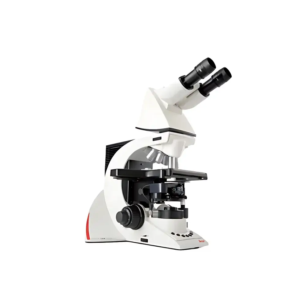
Leica DM3000 Intelligent Biological Microscope
| Brand | Leica |
|---|---|
| Origin | USA |
| Manufacturer Type | Authorized Distributor |
| Origin Category | Imported |
| Model | Leica DM3000 Intelligent Biological Microscope |
| Pricing | Upon Request |
Overview
The Leica DM3000 Intelligent Biological Microscope is an ergonomically engineered upright microscope designed for high-throughput, long-duration applications in clinical pathology, cytology, hematology, and biomedical research. Built upon Leica’s proven infinity-corrected optical architecture, the DM3000 delivers consistent, high-contrast, aberration-corrected imaging across all standard contrast modes—including brightfield, darkfield, phase contrast, polarized light, and fluorescence. Its core intelligence lies in adaptive illumination management and motorized optical coordination: the system automatically identifies objective magnification via encoder feedback from the 6-position motorized nosepiece, then dynamically adjusts condenser top-lens position (automatically engaging or retracting the swing-out top lens for objectives ≤10×) and halogen/LED light intensity to match pre-stored user-defined illumination profiles. This eliminates manual reconfiguration between magnifications—reducing operator fatigue during extended diagnostic sessions and improving inter-observer consistency in clinical workflows.
Key Features
- Infinity-corrected optical path with optional Plan Achromat, Plan Semi-Apochromat, or Plan Apochromat objectives for optimal flatness and chromatic correction
- Motorized 6-position objective turret with encoder-based magnification recognition
- Motorized condenser with automatic top-lens swing-in/swing-out for objectives ≤10×—ensuring optimal numerical aperture matching without manual intervention
- Intelligent illumination system: 12 V / 30 W halogen or high-CRI LED source with automatic brightness recall per objective; supports dual-objective Toggle mode for rapid comparison between two user-selected magnifications
- Ergonomic design: reversible focus knobs, one-handed specimen stage control with low-profile mechanical stage, and ten programmable front-panel shortcut buttons—including dedicated Toggle and turret control keys
- Flexible imaging integration: C-mount port compatible with CCD/CMOS cameras; supports third-party acquisition software (e.g., LAS X, NIS-Elements, ImageJ plugins) and DICOM-compliant digital pathology workflows
- Expandable modular platform: supports multi-user viewing heads, drawing tubes, macro modules, and footswitch-controlled objective switching for hands-free operation
Sample Compatibility & Compliance
The DM3000 accommodates standard glass slides (1 × 3 inches), Petri dishes, culture flasks, and tissue sections mounted on conventional histological substrates. Its mechanical stage offers precise X–Y translation (76 × 52 mm travel) with fine-tuned vernier scales and optional encoded positioning for reproducible region-of-interest navigation. The system complies with IEC 61000-4 electromagnetic compatibility standards and meets EN 61326-1 requirements for laboratory equipment. When configured with audit-trail-enabled imaging software and user-access controls, the platform supports GLP/GMP-aligned documentation practices and aligns with FDA 21 CFR Part 11 principles for electronic records and signatures in regulated environments. Optional fluorescence filter cubes (5-position turret) conform to ISO 8578:2017 spectral transmission specifications for diagnostic fluorescence microscopy.
Software & Data Management
The DM3000 interfaces seamlessly with Leica Application Suite (LAS) X and third-party acquisition platforms via USB 2.0 or GigE Vision protocols. All illumination and turret settings are stored in non-volatile memory per user profile—enabling lab-wide standardization of imaging parameters. Metadata (objective ID, condenser position, light intensity, exposure time) is embedded in TIFF/OME-TIFF image headers, facilitating traceability and downstream analysis in LIMS or digital pathology archives. Optional LAS X modules provide measurement tools compliant with ASTM E1558 (quantitative metallography) and ISO 9276-2 (particle size distribution analysis), supporting QC/QA reporting in academic and clinical laboratories.
Applications
- Clinical pathology: Routine H&E, Pap smear, bone marrow aspirate, and peripheral blood film evaluation under standardized illumination conditions
- Academic research: Live-cell observation (with environmental chamber compatibility), immunofluorescence co-localization, and morphometric analysis of cultured neurons or stem cell colonies
- Teaching laboratories: Multi-station configurations enable synchronized instruction and real-time annotation sharing across networked workstations
- Digital pathology pre-screening: Integration with whole-slide scanning systems for rapid triage and ROI selection prior to high-resolution digitization
- Hematology: Differential white blood cell counts, platelet morphology assessment, and reticulocyte enumeration using optimized phase contrast and brightfield protocols
FAQ
Does the DM3000 support fluorescence imaging out of the box?
Yes—the system includes a five-position filter cube turret and accepts standard Leica fluorescence cubes (e.g., GFP, TRITC, DAPI). Excitation/emission spectra comply with ISO 8578:2017 for clinical fluorescence applications.
Can illumination profiles be exported or backed up across multiple instruments?
Yes—user profiles including illumination settings, turret assignments, and shortcut configurations can be saved as XML files and deployed across other DM3000 units within the same facility.
Is the footswitch interface compatible with third-party peripherals?
The DM3000 uses a standard 3.5 mm mono jack input; it is compatible with most commercially available momentary-contact footswitches meeting IEC 60601-1 safety requirements for medical electrical equipment.
What is the maximum field number supported by the eyepieces?
Standard 10× wide-field eyepieces support a 23 mm field number; optional 10× eyepieces with 25 mm field number are available for expanded viewing area without compromising edge-to-edge resolution.
How does the automatic condenser top-lens mechanism improve quantitative reproducibility?
By eliminating manual condenser alignment variability, the motorized top-lens ensures consistent Köhler illumination geometry across users and sessions—critical for comparative morphometry and automated image analysis pipelines.


