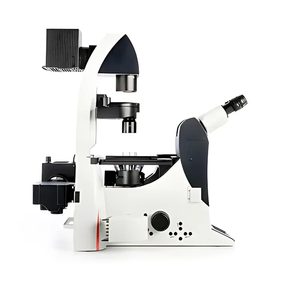
Leica DMI4000 / DMI6000 Fully Automated Inverted Biological Microscope
| Brand | Leica |
|---|---|
| Origin | Germany |
| Model | DMI4000 / DMI6000 |
| Type | Fully Automated Inverted Microscope for Live-Cell Imaging |
| Motorized Functions | Motorized Focus, Motorized Condenser (1–70 mm range), Motorized Filter Cube Turret (6-position), Motorized Field Diaphragm (Circular & Rectangular), Motorized Fluorescence Intensity Control (5-step), Motorized Z-Drive with Parfocal Stability |
| Optical Path | 4-output CCD port, 1.5× built-in optovar, 25 mm field number |
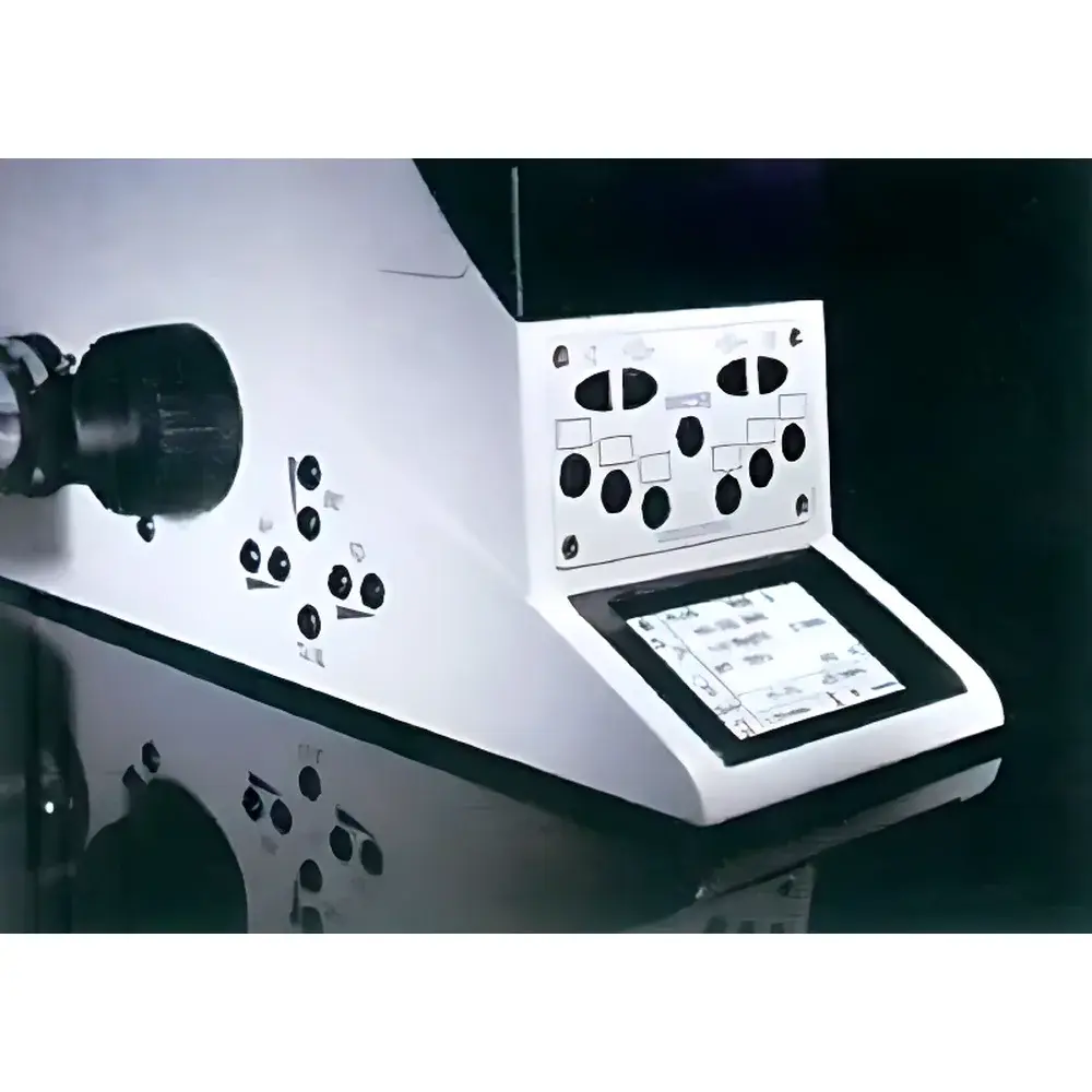
| Display | High-resolution parameter LCD with real-time error detection and visual alert feedback |
| Fluorescence Technology | Zero-Pixel-Drift Imaging, Stray-Light Trapping, Simultaneous FL/PH and FL/DIC multimodal acquisition |
| Compliance | Designed for GLP/GMP-aligned workflows |
Overview
The Leica DMI4000 and DMI6000 are fully automated inverted biological microscopes engineered for high-precision live-cell imaging, time-lapse microscopy, and quantitative fluorescence analysis in demanding research and clinical laboratory environments. Based on a rigid, vibration-damped optical architecture and precision-machined mechanical stage, these systems implement Couette-flow-stabilized Z-axis mechanics and motorized parfocal alignment to eliminate focus drift during extended acquisitions—critical for longitudinal studies of dynamic cellular processes. The core optical design follows Köhler illumination principles across all contrast modes, with seamless switching between brightfield, phase contrast (PH), differential interference contrast (DIC), and epifluorescence (FL) via a unified objective lens set. Unlike conventional inverted platforms requiring manual reconfiguration, the DMI series integrates hardware-level synchronization between condenser aperture, excitation filter selection, fluorescence intensity modulation, and camera output routing—enabling reproducible, operator-independent imaging protocols.
Key Features
- Fully motorized optical train: 6-position high-speed fluorescence filter cube turret (switching time <300 ms), motorized field diaphragm with selectable circular or rectangular apertures, and programmable 5-step LED or arc-lamp intensity control to minimize phototoxicity and photobleaching.
- Intelligent condenser system: 1–70 mm motorized height adjustment with automatic aperture matching to objective magnification and contrast mode; light intensity is memorized per configuration and restored upon recall.
- Z-axis stability architecture: Motorized coarse/fine focus with active parfocal compensation ensures zero Z-drift over multi-hour acquisitions—validated under ISO 9022-18 environmental testing conditions.
- Multimodal imaging capability: Single-objective compatibility across transmission (brightfield, PH, DIC) and epi-illumination (FL) modalities; one-click FL/PH or FL/DIC co-acquisition without optical realignment.
- Dual-output flexibility: Four independent camera ports (including two side ports and two rear ports), combined with a 1.5× internal optovar and 25 mm field number eyepiece optics, support simultaneous multi-sensor capture (e.g., sCMOS + EMCCD) and wide-field tiling.
- Human-centered interface: 25 programmable softkeys (DMI6000) with context-sensitive labeling; high-contrast LCD display with real-time parameter validation and visual error signaling (e.g., mismatched NA/condenser setting triggers persistent blink until correction).
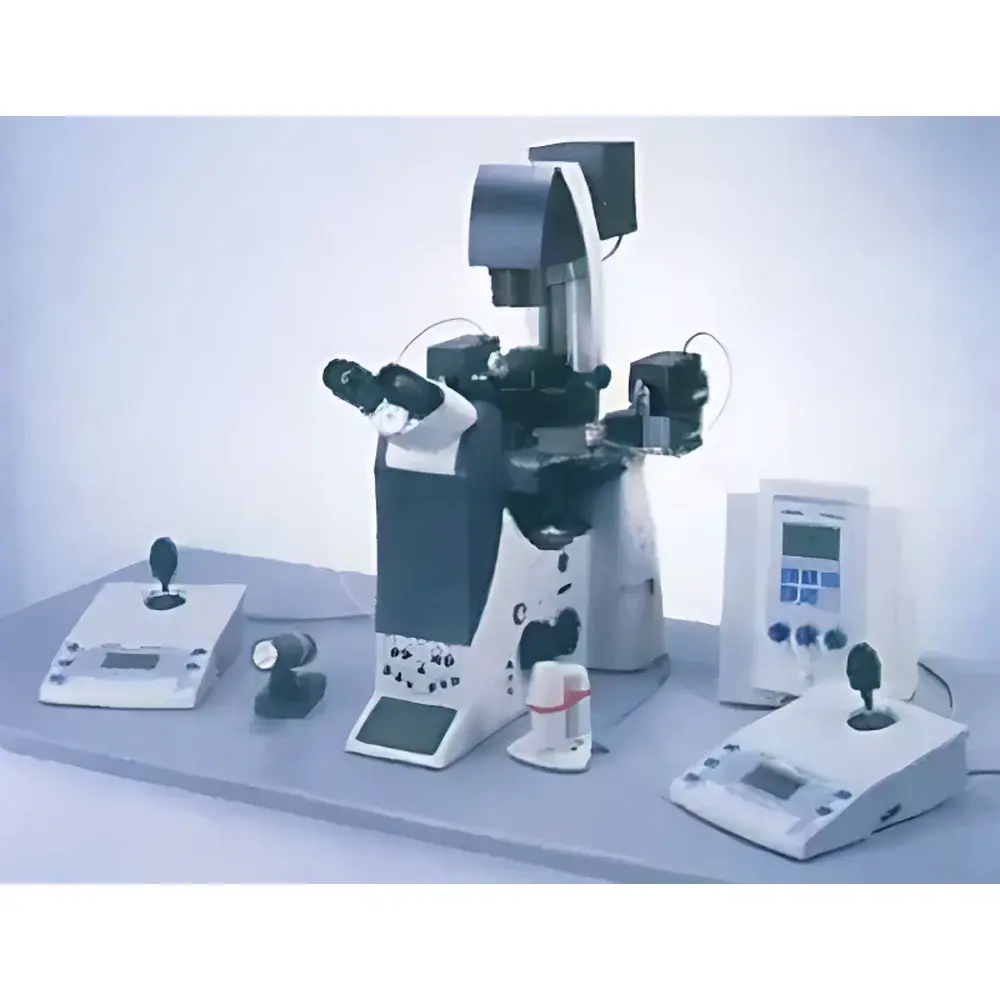
Sample Compatibility & Compliance
The DMI4000/DMI6000 platform accommodates standard and custom culture formats including 35 mm glass-bottom dishes, 6–96-well plates, microfluidic chambers, and perfusion-compatible stage-top incubators (e.g., Tokai Hit INU series). Its modular stage design supports motorized XY translation (optional) and precise thermal regulation interfaces. All fluorescence components—including dichroic mirrors, emission filters, and excitation sources—are calibrated per ISO 10110-7 surface quality standards and certified for spectral transmission consistency (±1.5 nm center wavelength tolerance). The system meets IEC 61000-6-3 EMC immunity requirements and is CE-marked for in vitro diagnostic (IVD) use where integrated with validated image analysis software. When deployed with Leica LAS X software in audit-mode configuration, it satisfies FDA 21 CFR Part 11 electronic record and signature requirements, including user authentication, session logging, and immutable audit trails.
Software & Data Management
Leica LAS X is the native acquisition and analysis platform, supporting script-based protocol automation (Python API), multi-dimensional time-series registration, and GPU-accelerated deconvolution. The software enables synchronized hardware control across all motorized axes, exposure parameters, and illumination states—ensuring full traceability from raw pixel data to final quantification. Metadata embedding follows MIAME/MINSEQ-compliant schema, with export options for OME-TIFF, ND2, and HDF5 containers. Integration with third-party analysis environments (e.g., Fiji/ImageJ, MATLAB, Python scikit-image) is facilitated via standardized TIFF header tags and JSON-formatted acquisition logs. For regulated environments, optional LAS X LMS (Laboratory Management System) module provides role-based access control, electronic signatures, and 21 CFR Part 11-compliant change history tracking.
Applications
- Intracytoplasmic sperm injection (ICSI) workflow support: High-NA phase contrast optics enable real-time visualization of oocyte spindle integrity and pronuclear formation without phototoxic stress.
- Live-cell calcium dynamics: Dual-channel ratiometric imaging (e.g., Fura-2) with sub-second temporal resolution and zero-pixel-shift registration across excitation wavelengths.
- Förster resonance energy transfer (FRET) quantification: Precise spectral unmixing using motorized dual-bandpass filter sets and hardware-synchronized exposure timing to minimize crosstalk artifacts.
- Long-term cell migration and division tracking: Stable Z-focus maintenance over >24 h enables reliable lineage tracing in wound-healing assays and organoid development studies.
- Multi-parametric phenotypic screening: Concurrent acquisition of morphology (DIC), membrane potential (voltage-sensitive dyes), and nuclear localization (Hoechst/DAPI) within single experimental runs.
FAQ
What distinguishes the DMI6000 from the DMI4000?
The DMI6000 adds full Z-axis motorization with active parfocal stabilization, 25 programmable function keys, enhanced condenser automation (including automatic NA matching), and expanded software scripting capabilities via LAS X Advanced Edition.
Is the system compatible with environmental control chambers?
Yes—the stage design includes standardized mounting interfaces and cable management pathways for integration with commercial CO₂/O₂/temperature-controlled incubation systems (e.g., OkoLab, Tokai Hit, PeCon).
Can fluorescence filter cubes be customized for non-standard fluorophores?
Leica offers OEM-grade customization of dichroic mirrors and bandpass filters through its Optics Engineering Group, with delivery lead times aligned to ISO 10110 manufacturing schedules.
Does the system support super-resolution techniques such as TIRF or SIM?
While the DMI platform is not a dedicated super-resolution system, its rigid mechanical design, motorized TIRF arm option (available as add-on), and precise XYZ repeatability make it suitable as a foundation for structured illumination (SIM) and total internal reflection fluorescence (TIRF) configurations when paired with appropriate lasers and cameras.
How is calibration traceability maintained across imaging sessions?
Each motorized component is factory-calibrated against NIST-traceable interferometric standards; calibration data is embedded in firmware and automatically applied during startup—no user recalibration required under normal operating conditions.


