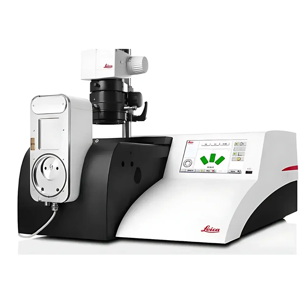
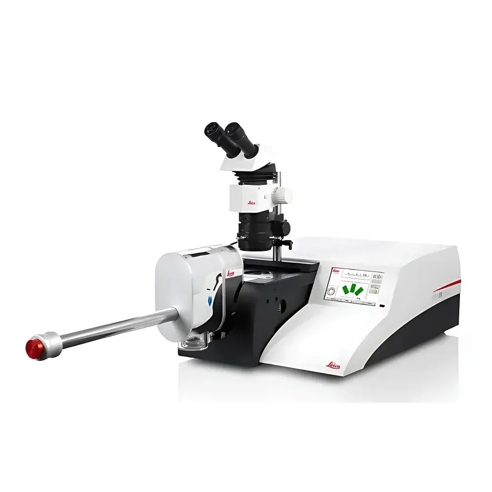
Leica EM TIC 3X Triple-Beam Ion Milling System
| Brand | Leica |
|---|---|
| Origin | Germany |
| Model | EM TIC 3X |
| Application | Research Use Only |
| Sample Throughput | Up to 3 specimens simultaneously |
| Beam Configuration | Three independently adjustable argon ion beams |
| Operating Temperature Range | Ambient to –120 °C (with optional cryo stage) |
| Compatible Interfaces | VCT vacuum transfer docking station, EM ACE600/ACE900 coaters, SEM systems |
Overview
The Leica EM TIC 3X is a high-precision triple-beam ion milling system engineered for the preparation of artifact-free cross-sections and ultra-smooth surfaces for advanced materials characterization. Utilizing three independently controllable argon ion beams operating in parallel, the system enables simultaneous slope cutting, planar polishing, and edge thinning—eliminating sequential processing steps and significantly reducing total preparation time. Unlike conventional single- or dual-beam ion mills, the EM TIC 3X’s tri-beam architecture delivers superior beam uniformity, minimized topographic shadowing, and enhanced control over milling angle and depth resolution—critical for preserving nanoscale structural fidelity in heterogeneous, multi-phase, or beam-sensitive specimens. Designed specifically for integration into end-to-end electron microscopy workflows, it supports both room-temperature and cryogenic operation (down to –120 °C), making it suitable for thermally labile polymers, hydrated biological tissues, soft composites, and geological samples containing volatile phases.
Key Features
- Triple argon ion beam configuration with independent angular adjustment (±15° to ±90°) for concurrent cross-sectioning and surface polishing
- Up to 2× faster material removal rate versus previous-generation ion mills, validated across metallic alloys, ceramics, and polymer blends
- Modular sample stage system supporting five interchangeable platforms: standard, multi-specimen (3-position), rotating, cryo-cooled (LN2-cooled), and vacuum-compatible VCT docking stage
- Integrated beam current monitoring and real-time ion dose logging for process reproducibility and traceability
- Automated beam alignment and endpoint detection via integrated secondary electron imaging (SEI) preview mode
- Full vacuum interlock compatibility with Leica EM ACE600/ACE900 sputter coaters and field-emission SEM platforms
Sample Compatibility & Compliance
The EM TIC 3X accommodates specimens up to 25 mm in diameter and 10 mm in height, including bulk metals, semiconductor wafers, fiber-reinforced composites, porous rocks (e.g., shale, carbonate), freeze-fractured biological tissue blocks, and water-soluble polymer fibers. Its cryo-capable configuration meets requirements for low-temperature TEM/SEM sample preparation under ISO 21748 (Guidance on uncertainty in quantitative microscopy) and ASTM E2931 (Standard Practice for Specimen Preparation for Scanning Electron Microscopy). When operated with the VCT docking interface and inert gas transfer modules, the system supports GLP-compliant workflow documentation—including timestamped beam parameter logs, stage position records, and vacuum history—for audit readiness in regulated R&D environments.
Software & Data Management
Controlled via Leica’s EM UC7-based GUI, the EM TIC 3X provides deterministic recipe management with version-controlled protocol storage, user-access-level permissions, and exportable XML-based method files. All operational parameters—including beam voltage (1–10 kV), current density (0.1–5 mA/cm²), milling duration, tilt angle, and cryo-stage temperature—are logged with UTC timestamps and linked to individual specimen IDs. Data exports comply with FAIR principles (Findable, Accessible, Interoperable, Reusable) and support integration into LIMS platforms via OPC UA or CSV batch export. Audit trail functionality satisfies FDA 21 CFR Part 11 requirements for electronic records and signatures when enabled with user authentication and electronic signature modules.
Applications
- Preparation of damage-free cross-sections for EBSD phase mapping in additively manufactured nickel superalloys
- Nanopore visualization in organic-rich shale matrices without hydrocarbon migration artifacts
- Interface analysis of multilayer thin-film stacks in photovoltaic devices (e.g., perovskite/SnO₂/TiO₂)
- Cryo-milled sections of vitrified plant root tissue for correlative light and electron microscopy (CLEM)
- Polishing of brittle intermetallic compounds for high-resolution EDS quantification without preferential sputtering
- Edge-thinning of battery electrode cross-sections (NMC/graphite) prior to FIB-SEM tomography
FAQ
What is the maximum specimen size supported by the EM TIC 3X?
The system accepts specimens up to 25 mm in diameter and 10 mm in height. Custom holders are available for irregular geometries under engineering consultation.
Can the EM TIC 3X be integrated with a focused ion beam (FIB-SEM) platform?
Yes—via the VCT vacuum transfer interface, specimens prepared in the EM TIC 3X can be transferred under <10⁻⁶ mbar to compatible FIB-SEM systems without air exposure.
Does the system support automated endpoint detection?
It includes SEI-based preview imaging for real-time visual monitoring; however, fully automated endpoint detection requires external integration with energy-filtered SEM or STEM detectors.
Is cryogenic operation validated for biological soft tissue?
Yes—validated protocols exist for plunge-frozen mammalian brain tissue and cryo-sectioned plant meristems, maintaining ice crystal dimensions below 50 nm per ISO 16700.
How is beam stability maintained during extended milling sessions?
The ion source incorporates active thermal regulation and closed-loop emission current feedback, ensuring ≤±1.5% beam current drift over 8-hour continuous operation.


