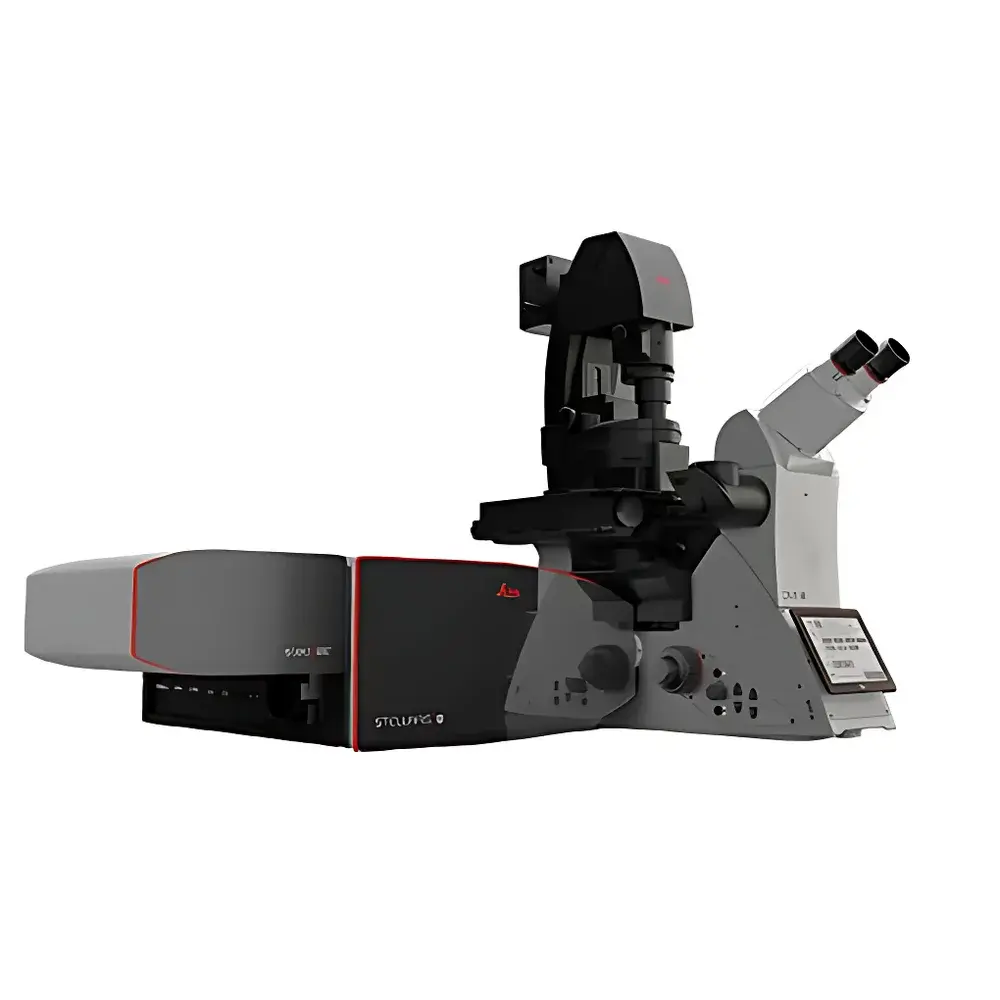
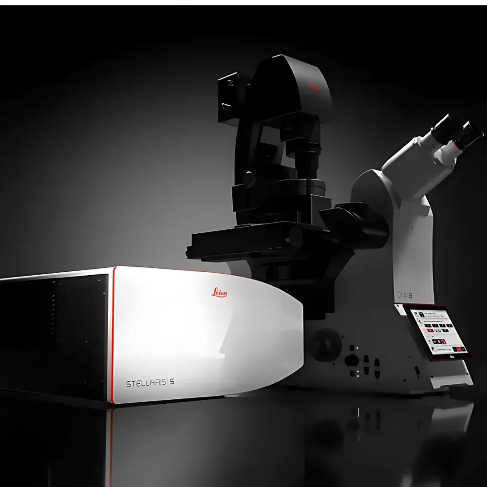
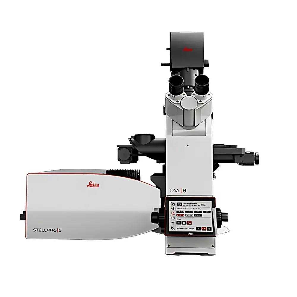
Leica STELLARIS Confocal Microscopy Platform
| Brand | Leica |
|---|---|
| Origin | Germany |
| Instrument Type | Point-Scanning Confocal Microscope |
| Laser Source | 8-line tunable white light laser (440–790 nm) |
| Detector | Power HyD hybrid detector series |
| Spectral Detection Range | 410–850 nm |
| Fluorescence Lifetime Imaging (FLIM) Capability | TauSense suite (TauContrast, TauGating, TauSeparation, TauInteraction) |
| Resolution Enhancement | LIGHTNING super-resolution engine with Dynamic Signal Enhancement (DSE) and AiviaMotion AI-accelerated real-time processing |
| Software Platform | LAS X Navigator + Aivia AI-powered analysis suite |
| Compliance | Designed for GLP/GMP-aligned workflows |
| Optical Architecture | Integrated acousto-optical beam splitter (AOBS), optimized light path, and photon arrival time digitization at pixel level (16 time-gated channels) |
Overview
The Leica STELLARIS Confocal Microscopy Platform represents a paradigm shift in high-content fluorescence imaging for life sciences. Engineered around the physical principle of time-resolved confocal detection, STELLARIS combines point-scanning architecture with single-photon timing resolution to extract fluorescence lifetime (τ) as an orthogonal contrast dimension—complementing spectral and spatial information. Unlike conventional spectral unmixing, which relies on emission wavelength separation, TauSense technology leverages nanosecond-scale decay kinetics to distinguish fluorophores with overlapping spectra, enabling robust multiplexing beyond the limits of conventional filter-based or spectral detection. The platform integrates a fully synchronized optical train: an 8-line tunable white light laser (WLL), AOBS-based excitation routing, and Power HyD hybrid detectors delivering photon detection efficiency (PDE) up to 58% in the visible range and triple that of PMTs in the NIR-I window (720–850 nm). This architecture supports quantitative, reproducible, and biologically gentle imaging—critical for longitudinal live-cell studies, organoid dynamics, and tissue-level functional mapping.
Key Features
- Power HyD hybrid detectors with >2× PDE vs. conventional PMTs (visible) and 3× improvement in NIR-I (720–850 nm), enabling high-fidelity detection of low-abundance probes and deep-tissue NIR markers
- TauSense FLIM suite: TauContrast for label-free functional readouts (e.g., metabolic state via NAD(P)H lifetime, pH via SNARF derivatives); TauGating for autofluorescence suppression and signal-to-background enhancement; TauSeparation for spectral overlap resolution without unmixing artifacts; TauInteraction for quantitative FRET/FLIM-FRET analysis
- LIGHTNING super-resolution engine: Real-time deconvolution with dynamic signal enhancement (DSE), delivering sub-diffraction lateral resolution (<140 nm) without compromising acquisition speed or photostability
- AiviaMotion: On-the-fly AI-driven motion correction and denoising, trained on biological ultrastructure priors, enabling stable imaging of beating cardiomyocytes or migrating neurons at full frame rate
- LAS X Navigator interface: Context-aware workflow guidance—from sample loading to publication-ready export—with automated parameter optimization based on objective, dye, and specimen type
- Modular expandability: Native compatibility with DIVE multiphoton, STED nanoscopy, DLS light-sheet, FALCON fast FLIM, and CARS vibrational imaging modules via shared optical backbone and unified software control
Sample Compatibility & Compliance
STELLARIS is validated for use across diverse biological specimens—including adherent and suspension mammalian cells, 3D organoids, zebrafish embryos, Drosophila tissues, and fixed human biopsy sections—without requiring specialized mounting media or refractive index matching beyond standard protocols. Its low-illumination-efficiency design minimizes phototoxicity, supporting >60-minute time-lapse imaging of sensitive primary neurons or stem cell colonies. From a regulatory standpoint, LAS X software complies with FDA 21 CFR Part 11 requirements for electronic records and signatures when configured with role-based access control, audit trail logging, and secure data encryption. All image metadata—including laser power, dwell time, pinhole size, detector gain, and τ-gating parameters—is embedded in OME-TIFF format per Open Microscopy Environment (OME) standards, ensuring traceability and interoperability with downstream analysis pipelines (e.g., QuPath, ilastik, Imaris). The system meets ISO 13485 design controls for research-use-only (RUO) instrumentation and supports GLP-compliant experiment documentation through customizable report templates.
Software & Data Management
LAS X Navigator serves as the central orchestration layer, abstracting hardware complexity while preserving full experimental control. Its “Intelligent Acquisition” mode recommends optimal settings based on fluorophore excitation/emission profiles and specimen thickness, reducing setup time by >70% for novice users. Aivia—Leica’s AI-native analysis engine—provides validated, containerized workflows for rare-event detection (e.g., mitotic errors, apoptotic bodies, metastatic protrusions), achieving >90% recall with <5% false positives on benchmark datasets. Aivia’s autonomous microscope mode triggers adaptive scanning only where biologically relevant events occur, cutting acquisition time by up to 70% and reducing raw data volume by 3–5× versus full-field capture. All processing occurs locally or on-premise HPC clusters; no cloud upload is required. Data export supports HDF5, N5, and OME-Zarr formats for scalable storage and integration with Python-based analysis ecosystems (e.g., Napari, scikit-image).
Applications
STELLARIS enables advanced applications spanning structural biology, neurodevelopment, immunology, and cancer metabolism. In live-cell metabolism studies, TauContrast quantifies mitochondrial membrane potential shifts via TMRM lifetime changes under hypoxic stress. In multiplexed tumor microenvironment profiling, TauSeparation resolves CD4/CD8/FOXP3 signals in FFPE sections despite spectral overlap from conventional Alexa Fluor conjugates. For synaptic plasticity research, LIGHTNING + AiviaMotion captures spine remodeling in real time with motion-corrected 3D stacks at 0.5 µm isotropic resolution. In developmental biology, combined DIVE multiphoton and STELLARIS FLIM reveal NADH/FAD⁺ redox ratios in gastrulating mouse embryos with cellular resolution. The platform also supports correlative workflows—e.g., sequential STELLARIS FLIM followed by cryo-EM grid preparation—via coordinate-mapped stage navigation.
FAQ
What distinguishes STELLARIS from legacy confocal platforms in terms of fluorescence lifetime imaging?
STELLARIS implements time-correlated single-photon counting (TCSPC) at the pixel level with 16 independently adjustable time gates, enabling true multi-parameter FLIM without scan-speed penalties or post-acquisition fitting artifacts.
Can STELLARIS be integrated into existing microscopy core facilities with mixed-brand infrastructure?
Yes—STELLARIS supports standardized communication protocols (Micro-Manager API, JSON-RPC over TCP/IP) and exports metadata-compliant OME-TIFF, ensuring seamless integration with facility-wide LIMS and data management systems.
Is TauSense compatible with common organic dyes and genetically encoded indicators?
Yes—TauContrast and TauSeparation are validated for >120 fluorophores including GFP/mCherry variants, Cy dyes, ATTO dyes, and small-molecule sensors (e.g., Fluo-4, BCECF), with lifetime calibration curves provided in the LAS X library.
Does STELLARIS support quantitative colocalization analysis incorporating lifetime data?
Yes—Aivia includes τ-weighted Pearson and Manders coefficients, enabling statistically rigorous colocalization assessment that accounts for both spatial proximity and molecular interaction states.
How does the Power HyD detector improve signal fidelity in thick tissue imaging?
Its high PDE across 410–850 nm, coupled with sub-nanosecond timing resolution and low dark count rate (<10 cps), preserves photon statistics even under low-signal conditions typical of scattering tissue—enabling reliable τ extraction at depths exceeding 100 µm in cleared samples.


