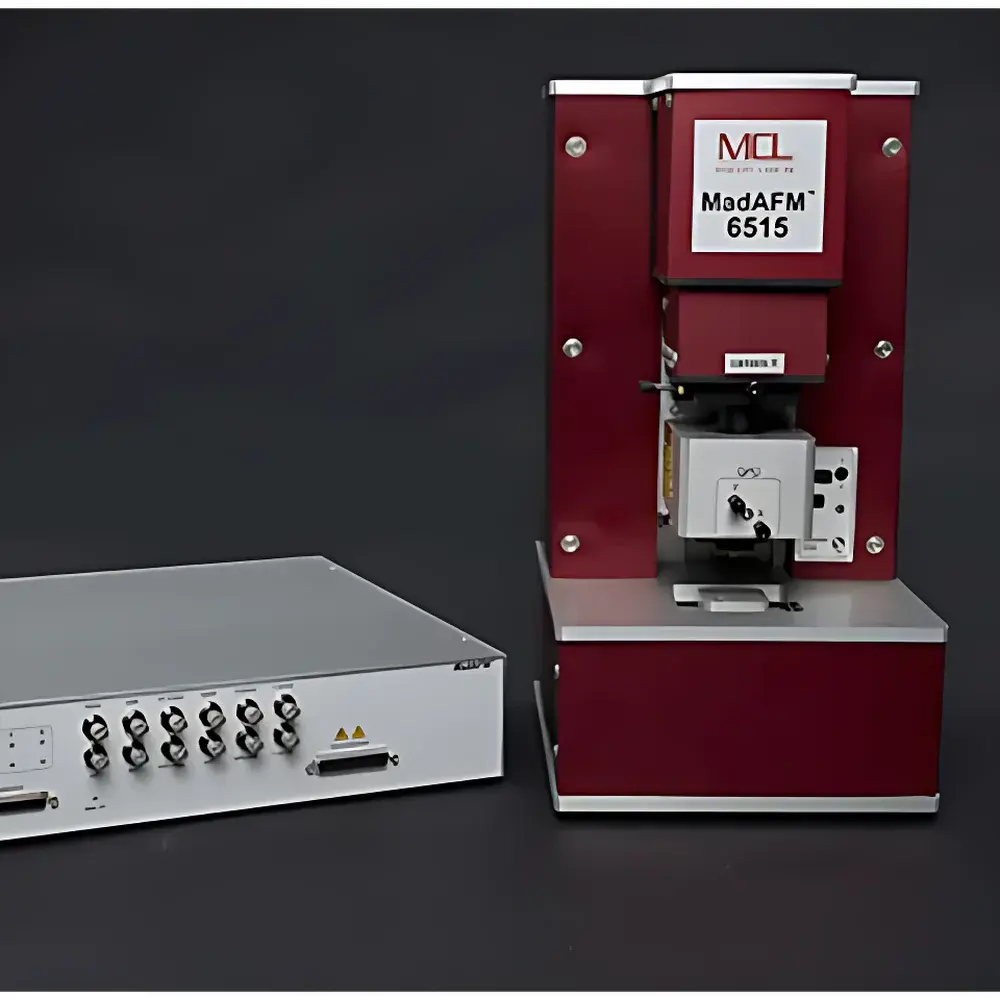
MadAFM™ Atomic Force Microscope by MCL Think Nano
| Brand | MCL Think Nano |
|---|---|
| Origin | USA |
| Model | MadAFM™ |
| Instrument Type | Material-Grade AFM |
| Positional Detection Noise | <0.15 nm RMS |
| Max Sample Size | 50 mm × 50 mm × 40 mm |
| XY Sample Stage Travel | 25 mm |
| Z Focus & Head Travel | 50 mm |
| Closed-Loop Nanopositioner Range (XY) | 30/65/100 µm |
| Z Nanopositioner Range | 15/30 µm |
| Nanopositioner Resolution (30 µm range) | 0.03 nm |
| Nanopositioner Noise Floor (peak-to-peak) | 2.7 pm |
| Optical Alignment | 635 nm Class II laser + 1.6 MP coaxial CMOS camera |
| Software | AFMView®-OD (USB 2.0 interface, Windows 8.1/10/11) |
Overview
The MadAFM™ Atomic Force Microscope by MCL Think Nano is a high-stability, desktop-compatible scanning probe microscope engineered for quantitative nanoscale surface characterization in academic, industrial, and quality control laboratories. Built upon over 25 years of proprietary piezoelectric nanopositioning expertise, the system employs a flexure-guided, closed-loop architecture with integrated PicoQ® low-noise capacitive sensors—delivering sub-picometer positional stability and <0.15 nm RMS detection noise in operational conditions. Unlike open-loop or hybrid-stage systems, the MadAFM™ implements full XYZ closed-loop control at the nanopositioner level, ensuring traceable, reproducible displacement without hysteresis or creep. Its measurement principle relies on force feedback between a sharp microfabricated cantilever and the sample surface, transduced via optical beam deflection (OBD) and processed in real time using a dual-phase lock-in amplifier. The instrument supports both contact and dynamic modes—including intermittent (tapping), non-contact, constant-height, and lateral force imaging—enabling topographic, mechanical, electrical, magnetic, and electrochemical property mapping across conductive, semiconductive, and insulating materials.
Key Features
- Proprietary PicoQ® capacitive sensors integrated into all three axes (X, Y, Z) of the nanopositioner, achieving 2.7 pm peak-to-peak noise floor and 0.03 nm step resolution over 30 µm travel range
- Flexure-guided, monolithic piezo nanopositioner design with <1 nm out-of-plane motion across full 100 µm scan range—critical for artifact-free phase and amplitude imaging
- Dual-stage motion architecture: high-resolution nanopositioner (30–100 µm XY, 15–30 µm Z) coupled with long-range motorized micropositioners (25 mm XY/focus, 50 mm Z), enabling seamless coarse-to-fine navigation
- Coaxial optical path with 1.6 MP CMOS camera, 85 mm focal length objective, and 635 nm alignment laser—providing real-time visual confirmation of cantilever–sample engagement and laser spot positioning
- Integrated white LED coaxial illumination and manual laser collimation—optimized for rapid setup and repeatability across diverse sample geometries and reflectivities
- Modular controller with 15-channel 24-bit ADC (dedicated to PicoQ sensors), 11-channel 20-bit DAC, and 14-channel I/O (12 BNC, including TTL triggers)—supporting synchronized external stimuli and multi-parameter acquisition
Sample Compatibility & Compliance
The MadAFM™ accommodates samples up to 50 mm × 50 mm × 40 mm and 500 g in mass, compatible with standard SEM stubs, silicon wafers, glass slides, polymer films, biological substrates (e.g., mica-supported lipid bilayers or fixed cells), and composite materials. Its rigid aluminum frame and optimized mass distribution minimize thermal drift and acoustic coupling, though optimal performance requires user-provided vibration isolation (e.g., pneumatic or active platforms). The system meets electromagnetic compatibility (EMC) requirements per FCC Part 15 Subpart B and CE Directive 2014/30/EU. While not certified for GMP environments, its hardware-level data integrity (full audit trail of DAC/ADC timestamps, sensor calibration logs, and parameter snapshots) supports GLP-compliant workflows. AFMView®-OD software stores raw time-series and metadata in HDF5 format, facilitating traceability per ISO/IEC 17025 and ASTM E2500-21 guidelines for instrument qualification.
Software & Data Management
AFMView®-OD serves as the native control and acquisition platform, offering automated initialization routines—including laser alignment verification, photodiode offset calibration, and cantilever resonance tuning—that reduce operator dependency and accelerate ramp-up for new users. Advanced users retain direct access to PID gains, feedback loop bandwidth, scan speed, setpoint modulation, and trigger timing—without requiring source-code modification. All acquired data are timestamped, sensor-calibrated, and stored with embedded metadata (e.g., scanner voltage maps, photodiode gain settings, environmental log headers). Export options include ASCII, Gwyddion (.gwy), MountainsSPIP® (.sur/.msr), and MATLAB-compatible .mat formats. The software supports USB 2.0 communication with Windows 8.1/10/11 (32/64-bit), requiring ≥4 GB RAM and a 3.1 GHz CPU. No cloud connectivity or telemetry is implemented—ensuring full local data sovereignty.
Applications
- Topographic & Mechanical Mapping: Quantitative roughness analysis (Sa, Sq, Sz per ISO 25178), elastic modulus extraction via force-distance spectroscopy, and viscoelastic profiling using force modulation microscopy
- Electrical Characterization: Conductive AFM (C-AFM) for current mapping, Kelvin probe force microscopy (KPFM) for surface potential, piezoelectric force microscopy (PFM) for ferroelectric domain imaging, and I-V spectroscopy under bias
- Magnetic Imaging: Magnetic force microscopy (MFM) with phase-locked lift-mode operation for stray field visualization in thin-film media and spintronic devices
- Nanoscale Patterning: Dip-pen nanolithography and local oxidation lithography enabled by precise Z-axis force control and tip-sample distance regulation
- Biological & Soft Matter Studies: In-air and liquid-cell imaging (with optional Open/Closed Cell Kits) of proteins, DNA, vesicles, and hydrogels—leveraging low-force intermittent mode to preserve structural integrity
FAQ
Does the MadAFM™ support liquid-phase imaging?
Yes—via optional Open Cell Kit (for ambient liquid droplets) or Closed Cell Kit (for controlled fluid environment and temperature stabilization). Both kits maintain optical access and laser alignment integrity.
Is the system compatible with third-party probes?
Yes—the MadAFM™ uses standard 125 µm-wide, 45°-backside coated cantilevers (e.g., Bruker RTESPA, Nanoworld Arrow) and features tool-free, alignment-free probe mounting with integrated preload adjustment.
What is required for regulatory compliance in pharmaceutical QC labs?
While the hardware supports audit trails and calibrated position reporting, formal 21 CFR Part 11 compliance requires supplementary validation documentation, electronic signature modules, and IT infrastructure controls—available through MCL’s qualified installation services.
Can multiple imaging modes be run simultaneously?
Yes—phase and topography channels are acquired synchronously in tapping mode; KPFM and PFM require sequential lift-mode operation but share identical scanner trajectories and calibration references.
Is vibration isolation included with the system?
No—vibration and acoustic isolation must be provided separately. MCL recommends active platforms (e.g., Minus K or Accurion) or high-mass passive tables for sub-nanometer stability in non-basement lab environments.


