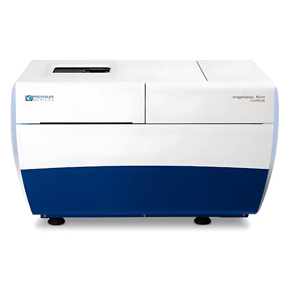
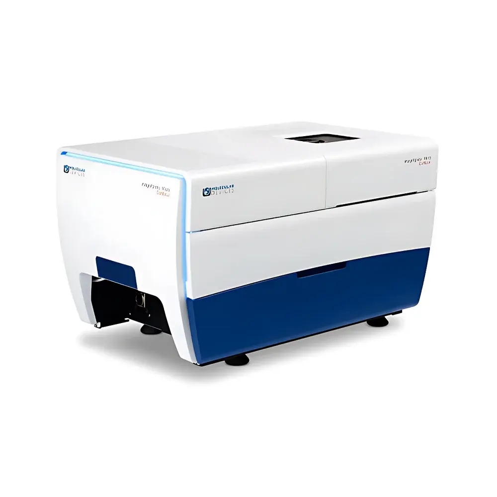
Molecular Devices ImageXpress Micro Confocal High-Content Imaging System
| Brand | Molecular Devices |
|---|---|
| Origin | USA |
| Manufacturer Type | Original Equipment Manufacturer (OEM) |
| Product Category | Imported Instrument |
| Model | ImageXpress Micro Confocal |
| Pricing | Available Upon Request |
Overview
The Molecular Devices ImageXpress Micro Confocal High-Content Imaging System is a fully automated, upright-style confocal imaging platform engineered for quantitative, multi-parametric analysis of fixed and live biological specimens across 2D monolayers, 3D cellular spheroids, organoids, tissue sections, and whole small model organisms (e.g., zebrafish embryos, C. elegans). Leveraging spinning-disk confocal microscopy with real-time hardware autofocus and software-driven Z-stack acquisition, the system eliminates out-of-focus blur inherent in widefield fluorescence imaging—enabling precise optical sectioning, high signal-to-noise ratio (SNR), and reproducible volumetric quantification. Its modular architecture supports seamless integration of environmental control (temperature, CO₂, humidity), transmitted-light contrast modes (phase contrast, DIC), and liquid handling peripherals—making it suitable for longitudinal assays requiring physiological relevance and statistical robustness. Designed for GLP-compliant labs and regulated research environments, the system adheres to core principles of assay traceability, metadata-rich image capture, and audit-ready data provenance.
Key Features
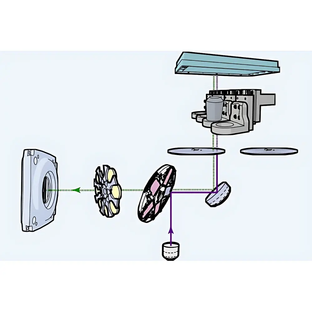
- AgileOptix™ Technology: One-button switching between widefield, confocal, and transmitted-light imaging modes without mechanical reconfiguration or alignment recalibration.
- High-Speed Spinning-Disk Confocal Engine: Delivers diffraction-limited resolution (lateral ~250 nm, axial ~600 nm) at acquisition speeds up to 30 fps per channel—maintaining throughput across 96-, 384-, and 1536-well microplates.
- Scientific CMOS Detector: Large-format sensor (≥2048 × 2048 pixels) with >3-log dynamic range, enabling simultaneous detection of low-abundance and saturated signals within a single exposure.
- Laser-Based Excitation Options: Configurable 5-channel (400–1000 mW/channel) or 8-channel (including NIR) solid-state laser module; enables multiplexed immunofluorescence, FRET, and deep-tissue excitation with reduced phototoxicity.
- Water-Immersion Objective Support: Compatible with high-NA water-dipping objectives (e.g., 20×/1.0 W, 40×/1.15 W); increases photon collection efficiency by up to 4× in thick samples, improves Z-resolution, and minimizes spherical aberration in Matrigel-embedded spheroids and brain slices.
- Hardware-Autofocus + Software Focus Mapping: Dual-layer focusing ensures uniform focus across entire wells—even on uneven substrates or curved tissue surfaces—critical for plate-wide consistency in phenotypic screening.
Sample Compatibility & Compliance
The system accommodates diverse specimen formats: standard flat-bottom, U-bottom, and hanging-drop microplates; glass- or polymer-based tissue culture inserts; formalin-fixed paraffin-embedded (FFPE) and cryosections (up to 200 µm thickness); and live specimens in custom chambers. Environmental control modules maintain physiological conditions (37°C ± 0.3°C, 5% CO₂, 95% RH) for ≥72-hour time-lapse imaging of stem cell differentiation, tumor invasion, or neurodevelopmental dynamics. All image metadata—including instrument configuration, exposure parameters, objective ID, and calibration timestamps—is embedded in TIFF headers and archived in MDCStore™, ensuring full compliance with FDA 21 CFR Part 11 requirements for electronic records and signatures when deployed with validated workflows.
Software & Data Management
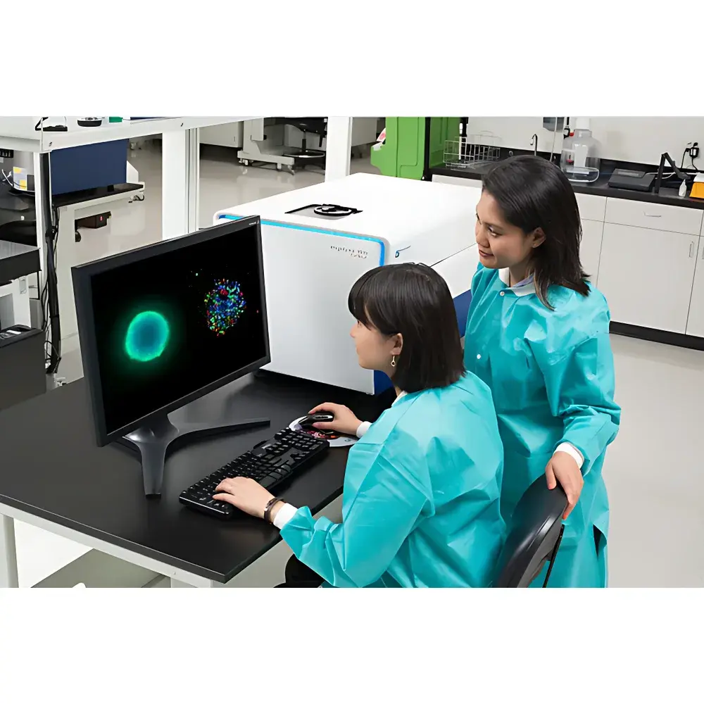
MetaXpress® High-Content Analysis Software provides an integrated environment for acquisition, segmentation, feature extraction, and statistical modeling—all within a single GUI. It includes over 120 pre-validated analysis modules (e.g., nuclear translocation, neurite outgrowth, mitochondrial morphology, mitotic index) and supports user-defined pipelines via the Custom Module Editor (CME), incorporating convolutional filters, machine-learning classifiers, and adaptive background correction (Adaptive Background Correction™). AcuityXpress™ enables database-backed statistical visualization—supporting dose-response curve fitting (4PL, Hill equation), PCA, hierarchical clustering, and export to third-party tools (R, Python, GraphPad Prism). All raw and processed data reside in the MDCStore™ relational database, which accepts external inputs from other imaging platforms (e.g., Zeiss LSM, Nikon A1R) and supports SQL-based querying, role-based access control, and automated backup policies.
Applications
The ImageXpress Micro Confocal system is routinely deployed in target identification and validation, lead optimization, toxicology assessment, and mechanistic pharmacology. Specific use cases include: quantitative analysis of 3D tumor spheroid viability and drug penetration kinetics; high-content profiling of iPSC-derived cardiomyocyte beating dynamics and calcium transients; spatial mapping of synaptic density in cortical organoids; longitudinal tracking of macrophage phagocytosis in co-culture models; and morphometric quantification of axon guidance defects in zebrafish neural tubes. Its ability to resolve subcellular features (e.g., puncta distribution, organelle fragmentation, chromatin condensation) while preserving spatial context makes it indispensable for translational biomarker discovery and functional genomics screening.
FAQ
Does the system support live-cell imaging under controlled environmental conditions?
Yes—optional environmental control units enable stable temperature (20–44°C), CO₂ (0–20%), and humidity regulation for long-term kinetic assays.
Can I import and analyze images acquired on other microscope platforms?
Yes—MDCStore™ accepts TIFF, OME-TIFF, and ND2 formats; MetaXpress® can process imported datasets using identical analysis modules and metadata tagging protocols.
Is the software compliant with FDA 21 CFR Part 11 for regulated biopharma workflows?
When deployed with validated installation qualification (IQ), operational qualification (OQ), and documented user training, the MetaXpress®/MDCStore™ suite supports electronic signature, audit trail, and role-based permissions required for GxP environments.
What Z-stack reconstruction and 3D rendering capabilities are available?
The system supports automated multi-plane acquisition, maximum-intensity projection (MIP), minimum-intensity projection, sum projection, and surface-rendered 3D volume visualization—exportable as STL or OBJ for downstream structural analysis.
How does the water-immersion objective improve imaging of thick 3D cultures?
By matching the refractive index of aqueous media, water-dipping objectives reduce spherical aberration and increase effective working distance—enhancing signal penetration depth, axial resolution, and quantitative accuracy in Matrigel-embedded or collagen-embedded models.


