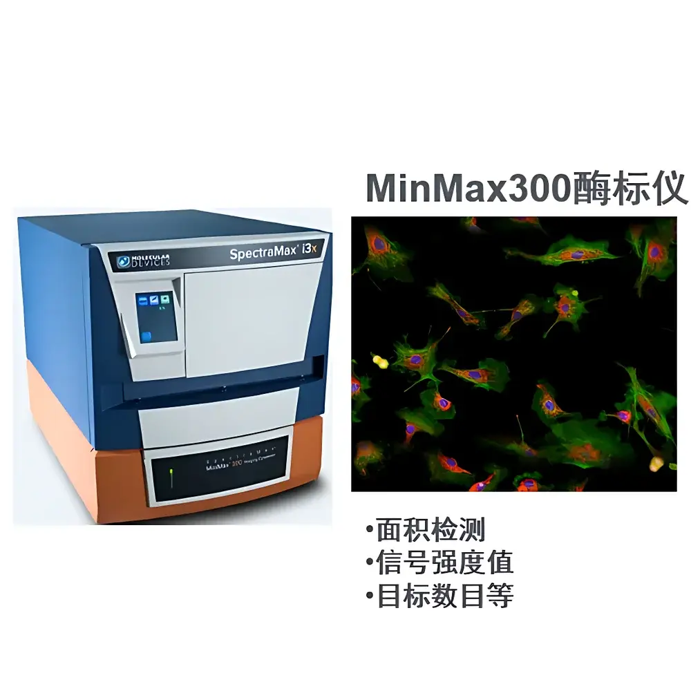
Molecular Devices SpectraMax MiniMax 300 Cell Imaging System
| Brand | Molecular Devices |
|---|---|
| Origin | USA |
| Manufacturer Type | Original Equipment Manufacturer (OEM) |
| Import Status | Imported |
| Model | SpectraMax MiniMax 300 Cell Imaging System |
| Instrument Type | Optical Imaging System |
| Compatibility | Designed exclusively for integration with SpectraMax i3x Multi-Mode Microplate Readers |
Overview
The Molecular Devices SpectraMax MiniMax 300 Cell Imaging System is a dedicated, modular optical imaging module engineered for seamless integration with the SpectraMax i3x multi-mode microplate reader platform. It transforms the i3x from a conventional plate reader into a hybrid instrument capable of quantitative fluorescence and brightfield cellular imaging—without requiring separate hardware, dedicated bench space, or complex infrastructure. The system operates on widefield optical imaging principles, leveraging high-resolution monochrome CMOS sensor technology coupled with precision motorized stage control and integrated LED-based excitation sources. Its core architecture supports label-free morphological analysis in transmitted light and dual-channel (green/red) epifluorescence detection, enabling concurrent functional assays and spatially resolved cell phenotyping within standard 6–384-well microplates. Designed for routine use in academic core facilities, pharmaceutical QC labs, and translational research environments, the MiniMax 300 adheres to fundamental requirements for reproducible, plate-based cellular imaging—including consistent illumination uniformity, calibrated pixel-to-micron mapping, and thermal stability during extended acquisition sequences.
Key Features
- Modular plug-and-play integration with SpectraMax i3x platforms—no external PC interface or standalone controller required
- Brightfield (transmitted light) imaging with proprietary label-free segmentation algorithms for confluence, cell count, and attachment quantification—eliminating reliance on viability dyes or fixation
- Dual-band fluorescence detection: optimized 470/40 nm (excitation) + 525/50 nm (emission) for GFP/YFP; 545/25 nm (excitation) + 605/55 nm (emission) for RFP/mCherry—configured via internal dichroic filters and LED switching
- Automated multi-field-of-view (FOV) acquisition per well: user-defined number of positions, z-stack capability (focus stepping), and adaptive exposure control
- Embedded image processing pipeline including background subtraction, contrast normalization, and sub-pixel centroid detection for single-cell object identification
Sample Compatibility & Compliance
The MiniMax 300 supports standard tissue-culture-treated and non-treated polystyrene microplates (6–384-well), including black-walled, clear-bottom formats essential for fluorescence and phase-contrast compatibility. It accommodates common cell types—including adherent lines (HEK293, HeLa, A549), primary neurons, and co-cultures—with no modification to culture protocols. All imaging workflows are compatible with GLP-aligned documentation practices: SoftMax Pro software maintains full audit trail functionality per FDA 21 CFR Part 11 requirements, including electronic signatures, session logging, and immutable data export (TIFF, PNG, CSV). The system complies with ISO/IEC 17025 traceability standards for measurement uncertainty reporting when used with calibrated reference plates (e.g., NIST-traceable fluorescent intensity standards).
Software & Data Management
Imaging acquisition, preprocessing, and quantitative analysis are fully managed through SoftMax Pro 7+ software, built upon the MetaMorph® imaging engine licensed from Molecular Devices. The interface provides wizard-driven protocol setup for plate mapping, channel selection, exposure optimization, and region-of-interest (ROI) definition. Built-in analysis modules include confluence estimation (based on pixel intensity thresholding and morphological erosion/dilation), fluorescent object counting (with size/shape gating), and ratiometric metrics (e.g., live/dead ratio, transfection efficiency %). Raw images are stored in vendor-neutral TIFF format with embedded EXIF metadata (exposure time, gain, LED intensity, objective magnification). Batch processing supports parallel analysis across multiple plates, with automated report generation compliant with SOP templates used in regulated biologics development.
Applications
- Label-free cytotoxicity screening: real-time monitoring of cell detachment and morphology changes induced by compound libraries
- Transfection efficiency quantification: co-localization analysis of fluorescent reporter constructs with nuclear stains (e.g., Hoechst 33342)
- Cell proliferation kinetics: longitudinal confluence tracking over 72–96 h without perturbing culture conditions
- Neurite outgrowth analysis: length, branching point, and process thickness measurements in primary neuronal cultures
- High-content validation of endpoint assays: orthogonal confirmation of ATP-based viability (CellTiter-Glo®) or caspase activation (Caspase-Glo®) results
FAQ
Can the MiniMax 300 be used independently of the SpectraMax i3x?
No—it is a hardware module designed exclusively for mechanical and electrical integration with the i3x platform. It lacks standalone power, cooling, or control interfaces.
Does the system support time-lapse imaging?
Yes—SoftMax Pro enables programmable interval acquisition across defined durations, with temperature and CO₂ control dependent on external incubation accessories connected to the i3x base unit.
What is the effective pixel resolution and field-of-view at 10× magnification?
The system delivers 1.2 µm/pixel sampling at 10× objective, yielding a 1.2 mm × 0.9 mm FOV per image—sufficient to resolve individual mammalian cells (10–20 µm diameter) with subcellular feature clarity.
Is third-party software integration supported?
Raw TIFF exports are fully compatible with ImageJ/Fiji, MATLAB, and Python-based analysis pipelines (e.g., scikit-image, CellProfiler); however, acquisition control and real-time hardware synchronization require SoftMax Pro.
How is focus maintained across multi-well plates with variable bottom thickness?
The system employs an auto-focus algorithm based on contrast maximization in brightfield mode, executed prior to each well’s acquisition sequence—compensating for ±0.1 mm variations in plate bottom geometry.
Related Products


