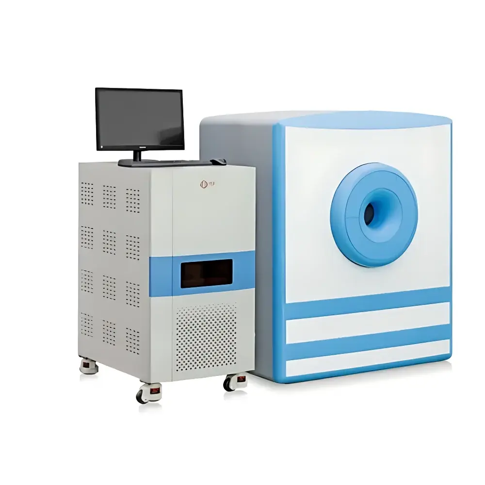
NIUMAG NM42-060H-I Preclinical MRI System for Glioma Animal Models
| Brand | NIUMAG |
|---|---|
| Origin | Jiangsu, China |
| Manufacturer Type | Authorized Distributor |
| Origin Category | Domestic (China) |
| Model | NM42-060H-I |
| Quotation | Upon Request |
| Magnetic Field Strength | 1.0 ± 0.05 T |
| Larmor Frequency | ~42 MHz |
| Dedicated Animal RF Coil Diameter | 60 mm |
| Species Compatibility | Live mice and rats |
| Primary Application | In vivo glioma modeling, tumor progression monitoring, therapeutic response assessment, contrast agent evaluation |
Overview
The NIUMAG NM42-060H-I is a dedicated 1.0 T preclinical magnetic resonance imaging (MRI) system engineered for longitudinal, non-invasive in vivo characterization of glioma animal models. Operating at a nominal Larmor frequency of 42 MHz for 1H nuclei, the system leverages permanent magnet architecture to deliver stable, low-drift field homogeneity—critical for quantitative T1, T2, and proton density-weighted imaging across repeated sessions. Unlike clinical or high-field research MRI platforms, the NM42-060H-I is purpose-built for rodent neuro-oncology studies: its 60 mm inner-diameter gradient-optimized RF coil ensures optimal signal-to-noise ratio (SNR) and spatial resolution within the murine cranium while maintaining physiological compatibility during extended anesthesia protocols. The system supports multi-parametric acquisition—including spin-echo, gradient-echo, and fast spin-echo sequences—enabling robust volumetric tumor segmentation, edema quantification, blood-brain barrier permeability assessment (via dynamic contrast-enhanced MRI), and treatment-induced necrosis mapping. Its design adheres to core principles of translational imaging: reproducibility across timepoints, compatibility with standard stereotactic surgery workflows, and integration with established glioma model systems (e.g., GL261 orthotopic, RCAS/tv-a transgenic, and lentiviral-driven PDGFB overexpression models).
Key Features
- Stable 1.0 ± 0.05 T permanent magnet with active shimming for field homogeneity ≤ 10 ppm over 30 mm DSV
- Dedicated 60 mm diameter transmit/receive volume coil optimized for murine brain imaging (FOV: 30 × 30 × 20 mm3)
- Integrated gradient subsystem with maximum strength ≥ 250 mT/m and slew rate ≥ 1,200 T/m/s for diffusion-weighted and functional MRI capability
- Real-time image reconstruction engine supporting on-board preview of axial, coronal, and sagittal slices within < 30 s post-acquisition
- Physiological monitoring interface for synchronized ECG gating, respiratory triggering, and temperature regulation (37.0 ± 0.3 °C)
- Modular software architecture compliant with DICOM 3.0 export and NIfTI-1 format for cross-platform analysis (FSL, SPM, ITK-SNAP)
Sample Compatibility & Compliance
The NM42-060H-I accommodates live C57BL/6, BALB/c, and nude mice (18–25 g) and Sprague-Dawley rats (200–250 g) under isoflurane anesthesia (1.5–2.5% v/v in O2). All animal handling procedures align with AAALAC International standards and EU Directive 2010/63/EU requirements for humane endpoints and imaging duration limits (< 90 min/session). The system’s RF exposure levels remain below IEC 60601-2-33 Class I safety thresholds for small-animal applications. Data acquisition workflows support ALARA (As Low As Reasonably Achievable) dose principles when used with gadolinium-based or iron oxide contrast agents, and all imaging protocols are documented per GLP Annex 11-compliant metadata tagging (including scanner parameters, animal ID, date/time, operator, anesthesia settings, and coil calibration logs).
Software & Data Management
NIUMAG’s proprietary MAGE-PRO v4.2 acquisition and analysis suite provides sequence programming via graphical pulse sequence editor (GPE), automated shimming routines, and ROI-based quantitative analysis modules for tumor volume, normalized enhancement ratio (NER), apparent diffusion coefficient (ADC), and T2 relaxation time mapping. Raw k-space data is stored in vendor-neutral HDF5 format with embedded BIDS-compatible sidecar JSON files. Audit trail functionality meets FDA 21 CFR Part 11 requirements: electronic signatures, immutable log entries, and role-based access control (RBAC) for protocol modification and report generation. Export pipelines integrate directly with MATLAB, Python (via nibabel and dcm2niix), and commercial platforms including Philips IntelliSpace Discovery and Siemens syngo.via.
Applications
- Orthotopic glioblastoma multiforme (GBM) model monitoring: volumetric growth kinetics, infiltrative margin delineation, and peritumoral edema evolution
- Evaluation of blood-brain barrier disruption using dynamic contrast-enhanced (DCE) MRI and Ktrans modeling
- Therapeutic response assessment following radiation, temozolomide, immune checkpoint inhibition, or oncolytic virotherapy
- Quantitative validation of novel MR contrast agents targeting EGFRvIII, integrin αvβ3, or macrophage infiltration
- Longitudinal metabolic phenotyping in obesity-associated glioma acceleration models (e.g., high-fat diet + RCAS-PDGFB)
- Cross-modal correlation with ex vivo histopathology (H&E, Ki-67, CD31), immunofluorescence, and spatial transcriptomics
FAQ
What is the minimum detectable glioma volume in mice using this system?
Typical in-plane resolution is 125 × 125 µm with 0.5 mm slice thickness; sub-2 mm3 lesions are reliably segmented using T2-weighted FLAIR and post-contrast T1-weighted sequences.
Can the system perform diffusion tensor imaging (DTI) in mouse brain?
Yes—b-values up to 2,000 s/mm2 and 32 diffusion-encoding directions are supported; FA and MD maps are generated natively in MAGE-PRO.
Is the magnet cryogen-free?
Yes—the 1.0 T permanent magnet requires no liquid helium or nitrogen; ambient temperature operation eliminates quench risk and reduces facility infrastructure demands.
Does the system support automated tumor segmentation?
MAGE-PRO includes semi-automated active contour (level-set) and deep learning-assisted (U-Net trained on annotated GL261 datasets) segmentation tools with inter-scan registration for longitudinal change detection.
How is compliance with Good Laboratory Practice (GLP) ensured during data acquisition?
All scans generate timestamped, digitally signed audit logs; raw data, parameter sets, and analysis scripts are archived with SHA-256 checksums and version-controlled metadata per OECD GLP Principles Section 5.2.
Related Products


