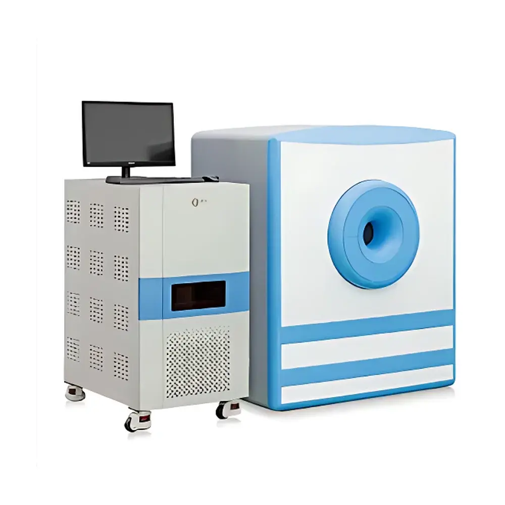
NIUMAG NM42 Low-Field Nuclear Magnetic Resonance Small Animal MRI System
| Brand | NIUMAG |
|---|---|
| Origin | Jiangsu, China |
| Magnet Type | Permanent Magnet |
| Field Strength | 1.0 T |
| Minimum Slice Thickness | 0.8 mm |
| Animal Weight Capacity | 1–45 g |
| Sample Diameter Limit | < 40 mm |
| Minimum Sample Volume | ≥ 100 µL |
| Scan Orientation | Arbitrary Angle & Arbitrary Plane |
| Compliance | Designed for Preclinical GLP-Compatible Workflow |
| Optional Modules | Gas Anesthesia System, ROI Analysis Software Suite |
Overview
The NIUMAG NM42 is a compact, permanent-magnet-based low-field nuclear magnetic resonance (NMR) imaging system engineered specifically for preclinical small animal research. Operating at a stable 1.0 Tesla field strength, the NM42 leverages optimized shimming and high homogeneity permanent magnet architecture to deliver robust signal-to-noise ratio (SNR) and spatial resolution—enabling reliable T1/T2 mapping, diffusion-weighted imaging, and quantitative relaxation analysis without cryogens or RF shielding infrastructure. Unlike high-field superconducting MRI systems, the NM42 eliminates the need for liquid helium, dedicated RF rooms, or structural reinforcement, making it suitable for standard laboratory environments—including shared core facilities and teaching labs. Its open-bore design accommodates live rodents up to 45 g with non-invasive, radiation-free longitudinal monitoring—ideal for pharmacokinetic studies, tumor progression tracking, and therapeutic response evaluation over multiple timepoints.
Key Features
- 1.0 T permanent magnet with passive shimming ensures field homogeneity ≤ 10 ppm over a 40 mm DSV (Diameter Spherical Volume), supporting reproducible quantitative NMR measurements and high-fidelity MRI reconstruction.
- Sub-millimeter imaging capability: Achieves isotropic in-plane resolution down to 80 µm with slice thickness adjustable to 0.8 mm across arbitrary oblique planes—facilitating precise anatomical localization in murine models.
- Non-invasive, non-ionizing operation: No ionizing radiation exposure; no tissue heating or acoustic noise above regulatory thresholds (IEC 60601-2-33 compliant design principles applied).
- Three-step automated acquisition workflow: Integrated auto-tuning, center-frequency search, and sequence parameter optimization reduce operator dependency—enabling consistent image quality even for users without formal MR physics training.
- Modular hardware expansion: Optional integrated gas anesthesia delivery (isoflurane/O2), physiological monitoring interface (respiratory gating, temperature feedback), and gradient coil upgrade kits support advanced functional and dynamic studies.
- Low total cost of ownership: Zero cryogen consumption; ambient-temperature operation; minimal power draw (< 1.5 kW); no RF cage or ferromagnetic exclusion zone required.
Sample Compatibility & Compliance
The NM42 accepts cylindrical samples with maximum diameter of 40 mm and minimum volume of 100 µL—compatible with standard NMR tubes (5 mm OD), microcentrifuge vials, and restrained live rodents (C57BL/6, BALB/c, nude mice, etc.). Animal positioning is facilitated by stereotactic holders with adjustable bite bars and ear pins. All pulse sequences adhere to IEC 62464-1 safety limits for specific absorption rate (SAR) and peripheral nerve stimulation (PNS). Data acquisition workflows are structured to support ALARA (As Low As Reasonably Achievable) principles and align with OECD Test Guidelines 407, 422, and 443 for repeat-dose toxicity studies. While not FDA-cleared as a diagnostic device, the system meets essential requirements for GLP-compliant preclinical data generation under ISO/IEC 17025:2017-accredited laboratories.
Software & Data Management
The NM42 runs on a Linux-based real-time acquisition platform with dual GUI layers: the core MRI console provides intuitive access to spin-echo, gradient-echo, inversion-recovery, and multi-echo CPMG sequences—with fully configurable TR/TE/TI, flip angle, bandwidth, and matrix size. Pulse timing parameters (RF pulse width, amplitude, gradient ramp time) are exposed via editable script templates, enabling method development for custom contrast mechanisms. The optional Image Processing Suite includes DICOM-compliant import/export, region-of-interest (ROI) quantification with statistical reporting (mean intensity, SD, CV%), multi-parametric color mapping (T2-weighted pseudocolor overlays), 3D volume rendering, and longitudinal change detection via rigid registration. Audit trails, user authentication, and electronic signature support comply with 21 CFR Part 11 requirements when deployed in regulated environments.
Applications
- Preclinical oncology: Longitudinal monitoring of xenograft tumor volume, necrosis fraction, and edema evolution using T2-weighted and contrast-enhanced T1 mapping.
- Contrast agent relaxivity characterization: In vitro r1/r2 determination per ISO 10993-18 standards; in vivo biodistribution kinetics via dynamic contrast-enhanced (DCE) MRI.
- Nanoparticle and colloidal dispersion analysis: Quantitative measurement of aggregation state, hydration shell thickness, and rotational correlation time via transverse relaxation dispersion (R2 dispersion) profiling.
- Metabolic phenotyping: Fat-water separation using Dixon-based techniques; hepatic steatosis grading in diet-induced obesity models.
- Neuroimaging: Ex vivo brain section imaging for infarct volume quantification post-MCAO; in vivo functional connectivity assessment via resting-state fMRI (rs-fMRI) with motion correction.
FAQ
What is the minimum animal weight supported by the NM42?
The system supports live rodent imaging from 1 g (e.g., neonatal mice) up to 45 g (e.g., mature Sprague-Dawley rats), provided the subject fits within the 40 mm transverse field-of-view.
Does the NM42 require an RF-shielded room?
No. The permanent magnet design and low operating frequency (~42.5 MHz at 1.0 T) eliminate the need for Faraday cages or specialized site preparation—installation only requires stable 230 V AC power and ambient temperature control (18–25 °C).
Can the NM42 perform quantitative T1 and T2 mapping?
Yes. Built-in inversion-recovery and multi-echo spin-echo sequences generate voxel-wise T1 and T2 maps with pixel-level uncertainty estimation, validated against ASTM E2857-13 reference phantoms.
Is anesthesia integration available as a factory option?
Yes. A CE-marked, flow-controlled isoflurane/O2 anesthesia module with scavenging interface and real-time vapor concentration feedback is available as an add-on configuration.
How is data security handled during acquisition and export?
All raw k-space data and reconstructed images are stored in vendor-neutral DICOM format with embedded metadata (pulse sequence, acquisition parameters, timestamp, operator ID); encrypted local storage and role-based access control are enabled via optional software license.


