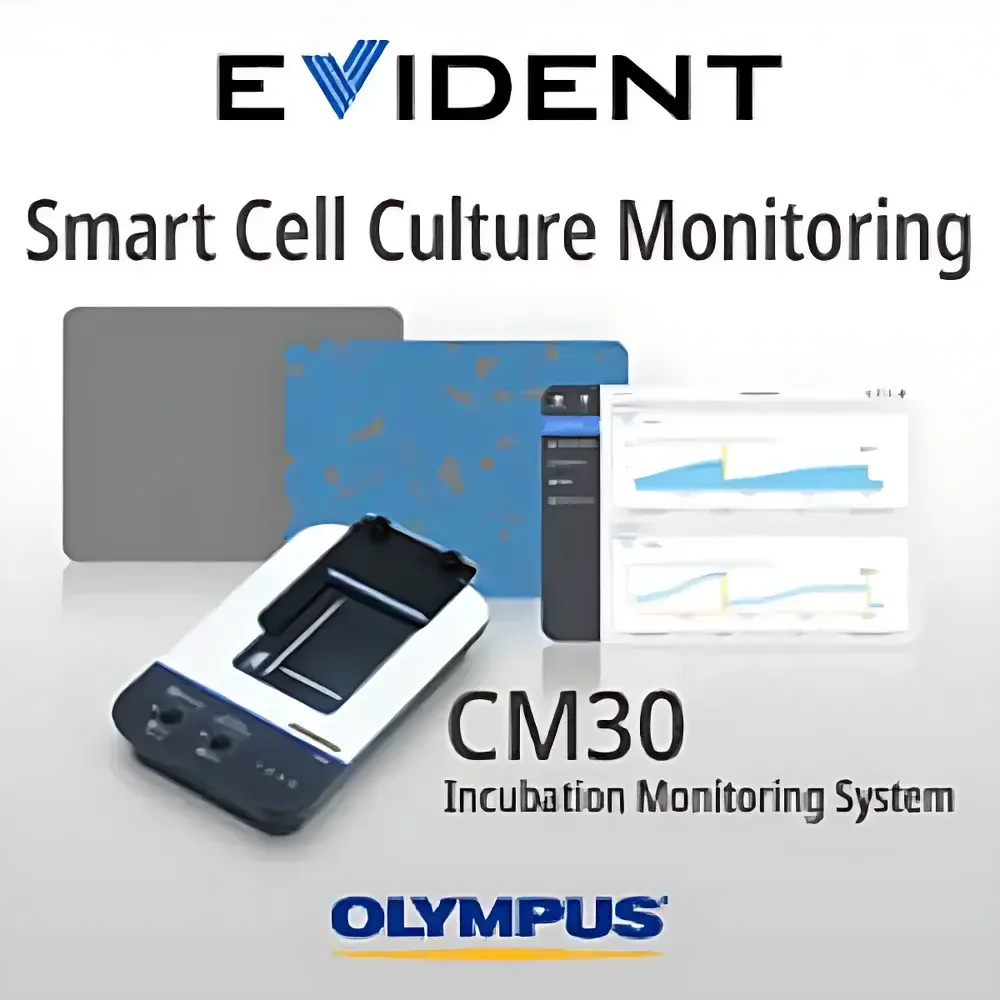
Olympus CM30 Cell Culture Monitoring System
| Brand | Olympus |
|---|---|
| Origin | Japan |
| Manufacturer | Olympus Corporation |
| Product Type | Imported Life Science Instrument |
| Model | CM30 |
| Pricing | Upon Request |
Overview
The Olympus CM30 Cell Culture Monitoring System is an integrated, incubator-embedded live-cell imaging platform engineered for label-free, non-invasive, longitudinal monitoring of adherent mammalian cells under physiologically relevant conditions. Unlike conventional inverted microscopes or standalone time-lapse systems, the CM30 operates autonomously inside standard CO₂ incubators (37 °C, 5% CO₂, >95% RH) without requiring user intervention or sample removal. Its core optical architecture employs 630 nm red LED illumination—selected to minimize phototoxicity and cellular stress—combined with oblique reflectance illumination (ORI), a proprietary contrast-enhancement technique that delivers high-contrast, pseudo-3D morphology visualization in brightfield mode. This enables reliable detection of subtle morphological dynamics—including mitosis, migration, confluence progression, and early-stage apoptosis—without fluorescent dyes or genetic labeling. The system captures quantitative image data at user-defined temporal intervals (e.g., every 15–120 minutes), feeding raw stacks into an embedded AI engine for real-time analysis and remote visualization via secure web interface.
Key Features
- Incubator-Integrated Design: Compact, fanless, and EMI-shielded housing certified for continuous operation inside Class II biosafety cabinets and standard humidified CO₂ incubators.
- Oblique Reflectance Illumination (ORI): Patented illumination geometry providing enhanced edge contrast and depth perception across diverse substrates—eliminating need for phase contrast, DIC, or Hoffman modulation optics.
- Motorized XYZ Stage: Precision stepper-driven stage with ±0.5 µm repeatability supports programmable multi-point acquisition across heterogeneous vessel formats (96-well plates, T25/T225 flasks, Petri dishes, cell factories, custom vessels).
- Auto-Focus Optimization: Adaptive focus algorithm calibrated for variable bottom thicknesses (0.12–2.0 mm) and refractive indices; supports user-defined vessel registration via intuitive calibration workflow.
- Large-Field Imaging: 2.84 mm × 2.13 mm field-of-view per frame (at 4× objective), enabling full-well or full-dish scanning with sub-minute acquisition time per position.
- On-Device AI Analytics: Edge-computing module trained on >10⁶ annotated cell images; performs real-time confluence quantification, single-cell counting, colony segmentation, and growth curve derivation without cloud dependency.
Sample Compatibility & Compliance
The CM30 is validated for use with standard tissue-culture-treated polystyrene (TCPS), glass-bottom, and hydrogel-coated substrates. It complies with ISO 13485:2016 (medical device quality management) and meets electromagnetic compatibility requirements per IEC 61326-1 for laboratory equipment. All firmware and software modules adhere to ALCOA+ principles for data integrity and are compatible with 21 CFR Part 11-compliant audit trail configurations when deployed in GMP-aligned workflows (e.g., monoclonal antibody process development, ATMP manufacturing). The system supports GLP documentation export including timestamped metadata, instrument logs, and raw TIFF stacks with embedded EXIF tags.
Software & Data Management
Olympus CM30 Control Suite (v3.2+) provides browser-based access (Chrome/Firefox/Edge) with role-based permissions (Admin, Operator, Viewer). Image data are stored locally on encrypted SSD with optional SFTP or HTTPS push to LIMS or ELN systems (e.g., LabVantage, Benchling). Each dataset includes machine-readable JSON metadata containing acquisition parameters, environmental sensor logs (temperature, CO₂, humidity), and AI confidence scores per analysis output. Batch processing pipelines support export of confluence heatmaps, population doubling time (PDT), scratch wound closure rates, and dose-response curves in CSV, PDF, or MIRIAM-compliant ISA-Tab format.
Applications
- Monoclonal antibody clonal expansion tracking and clone selection
- Stem cell passaging optimization and differentiation onset detection
- Primary cell viability assessment during cryopreservation recovery
- 3D spheroid size/growth kinetics monitoring in ultra-low attachment plates
- In vitro toxicology screening (IC₅₀ determination via confluence inhibition)
- Wound healing assay quantification (gap closure velocity, leading-edge dynamics)
- Bioprocess intermediate QC for cell therapy manufacturing (e.g., CAR-T expansion)
FAQ
Does the CM30 require external power or network cabling inside the incubator?
No—the CM30 uses a single PoE+ (IEEE 802.3at) connection for both power and Gigabit Ethernet communication, minimizing port penetration and maintaining incubator seal integrity.
Can the system be integrated with existing LIMS or MES platforms?
Yes—RESTful API and HL7-compatible webhooks enable bidirectional synchronization of sample IDs, acquisition schedules, and analytical results.
Is fluorescence imaging supported?
No—the CM30 is optimized exclusively for label-free brightfield ORI imaging; fluorescence capability is intentionally excluded to preserve long-term cell health and eliminate photobleaching artifacts.
What is the minimum detectable cell density for reliable AI counting?
Validation studies show ≥92% accuracy for confluent densities ≥5%, down to single isolated cells in low-density seeding conditions (e.g., limiting dilution assays).
How is data security ensured during remote access?
All communications use TLS 1.3 encryption; local storage employs AES-256 full-disk encryption; session timeouts and two-factor authentication (2FA) are configurable per organizational policy.


