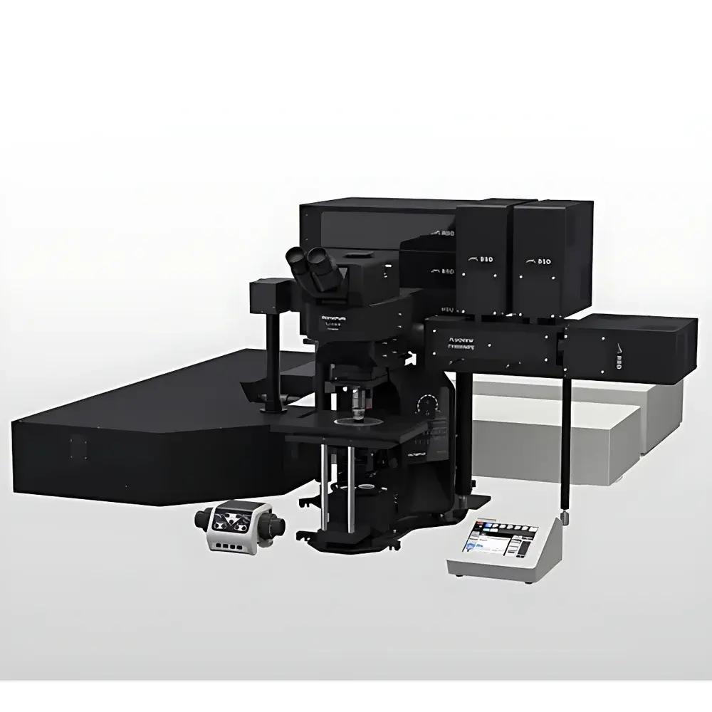
Olympus/EVIDENT FV4000MPE Multiphoton Laser Scanning Microscope
| Brand | Olympus/EVIDENT |
|---|---|
| Origin | Japan |
| Model | FV4000MPE |
| Instrument Type | Point-Scanning Multiphoton Confocal Microscope |
| Detector Technology | Silicon Photomultiplier (SiPM)-Based SilVIR Detectors |
| Maximum Detection Channels | 6 |
| Objective Compatibility | TruResolution Objectives |
| Scan Mode | Resonant Galvo Scanning |
| Application Focus | Live-Tissue & Deep-Tissue Multiphoton Imaging |
Overview
The Olympus/EVIDENT FV4000MPE Multiphoton Laser Scanning Microscope is an advanced point-scanning platform engineered for high-fidelity, deep-tissue fluorescence imaging in live biological specimens. It operates on the principle of multiphoton excitation—primarily two-photon and three-photon absorption—where near-infrared (NIR) pulsed lasers induce localized fluorophore excitation only at the focal plane, minimizing out-of-focus photodamage and enabling superior optical sectioning in scattering tissues up to several hundred micrometers in depth. Unlike conventional confocal systems relying on pinhole-based rejection of out-of-focus light, the FV4000MPE leverages intrinsic spatial confinement of multiphoton excitation, delivering inherently higher signal-to-background ratios and reduced phototoxicity during longitudinal live-cell and in vivo experiments. Its architecture integrates a modular optical path, tunable ultrafast laser coupling, and synchronized detection optimized for quantitative fluorescence intensity, lifetime, and spectral unmixing across multiple emission bands.
Key Features
- Patented SilVIR detector technology based on silicon photomultipliers (SiPM), offering high quantum efficiency (>40% at 500–900 nm), ultra-low read noise (<0.1 e⁻ RMS), and extended dynamic range (up to 6 decades), ensuring accurate quantification across weak and saturated signals.
- Up to six independent non-descanned detection (NDD) channels with individual gain, offset, and spectral filtering control—enabling simultaneous multicolor multiphoton acquisition without spectral crosstalk or mechanical filter wheel latency.
- TruResolution objective integration with adaptive spherical aberration correction, optimized for NIR transmission and high numerical aperture (NA ≥ 1.05) to maximize photon collection from deep tissue layers.
- Resonant galvanometric scanning unit capable of frame rates up to 30 fps at 1024 × 1024 pixels and line rates exceeding 30 kHz—supporting rapid volumetric time-lapse imaging of neuronal activity, calcium dynamics, and vascular perfusion.
- Modular beam path design compliant with standard OEM laser interfaces (e.g., Coherent Chameleon Discovery, Spectra-Physics Mai Tai HP), allowing seamless integration of dual-wavelength or OPO-based excitation sources for multimodal contrast generation.
Sample Compatibility & Compliance
The FV4000MPE supports a broad range of specimen formats including cultured primary neurons, brain slices (acute and organotypic), zebrafish embryos, mouse cortical windows, and explanted human tissue biopsies. Its low-phototoxicity imaging paradigm aligns with GLP-compliant longitudinal studies requiring repeated measurements over hours or days. The system meets ISO 13485 design control requirements for research-use-only instrumentation and supports audit-ready metadata logging per FDA 21 CFR Part 11 when paired with EVIDENT’s FV software v5.2 or later. All optical components comply with IEC 60825-1:2014 Class 4 laser safety standards, with integrated interlocks, shutter controls, and real-time power monitoring.
Software & Data Management
FV software provides a unified environment for acquisition, processing, and analysis—including batch deconvolution, Z-stack alignment, drift correction, and pixel-intensity calibration traceability. Raw data is saved in standardized OME-TIFF format with embedded metadata (excitation wavelength, PMT voltage, dwell time, stage coordinates). Time-series datasets support HDF5 export for interoperability with Python-based analysis pipelines (e.g., CaImAn, Suite2p). Audit trails record user actions, parameter changes, and instrument state transitions—essential for regulatory submissions under GxP frameworks. Optional FV Connect module enables remote monitoring and collaborative annotation via secure HTTPS endpoints.
Applications
- In vivo functional neuroimaging: Calcium dynamics in dendritic spines, microglial motility, and astrocytic Ca²⁺ waves in awake, head-fixed mice.
- Developmental biology: Cell migration tracking and morphogen gradient mapping in thick embryonic tissues using spectrally distinct fluorophores.
- Oncology research: Tumor microenvironment visualization—including immune cell infiltration, angiogenesis, and hypoxia sensing—within orthotopic xenograft models.
- Fibrosis and inflammation studies: Collagen SHG/THG contrast combined with immunofluorescence labeling in liver, lung, and kidney tissue sections.
- Pharmacokinetic profiling: Real-time drug distribution and metabolite formation in perfused organ-on-chip platforms.
FAQ
What laser sources are compatible with the FV4000MPE?
The system accepts Ti:Sapphire, OPO, and fiber-based ultrafast lasers with pulse widths <150 fs and repetition rates between 80–100 MHz. Standard coupling supports wavelengths from 680–1300 nm.
Can the FV4000MPE perform FLIM or spectral unmixing?
Yes—when equipped with time-correlated single-photon counting (TCSPC) hardware and spectral detectors, it supports fluorescence lifetime imaging microscopy (FLIM) and linear unmixing of >4 spectrally overlapping fluorophores.
Is the SilVIR detector cooled, and what is its typical dark count rate?
SilVIR detectors operate at thermoelectrically stabilized temperatures (−15°C), achieving dark count rates <500 cps/mm² at full gain—critical for low-light deep-tissue signal recovery.
How does the FV4000MPE handle chromatic dispersion across multiple excitation wavelengths?
TruResolution objectives incorporate multi-element achromatic correction optimized for NIR, while the scan lens and tube lens assembly include dispersion-compensating elements to maintain diffraction-limited focus across 700–1100 nm.
Does the system support automated tile scanning and stitching for large-area imaging?
Yes—motorized XY stages and piezo Z-drives integrate with FV software for programmable mosaic acquisition, with real-time overlap correction and seam-free stitching using phase-correlation algorithms.


