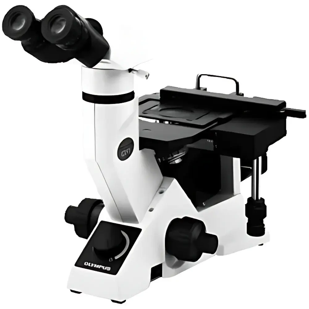
Olympus GX41 Inverted Metallurgical Microscope
| Brand | Olympus |
|---|---|
| Origin | Japan |
| Manufacturer | Olympus Corporation |
| Product Type | Inverted |
| Imaging System | Integrated digital imaging and analysis software |
| Total Magnification Range | 50×–600× |
| Eyepieces | Two (standard WHN10×, field number 22) |
| Objectives | Five UIS2 infinity-corrected objectives (5×, 10×, 20×, 50×, 100×) |
| Illumination | 6 V/30 W halogen or optional 100 W fiber-optic illuminator |
| Optical System | UIS2 infinity-corrected optics with flat-field plan apochromatic correction |
| Viewing Head | Tilting binocular tube (adjustable inclination for ergonomic standing operation) |
| Compliance | Designed to support ISO 9001-compliant quality control workflows |
Overview
The Olympus GX41 is a discontinued yet technically robust inverted metallurgical microscope engineered for high-reproducibility microstructural evaluation in industrial quality control, failure analysis, and materials science education environments. Its inverted optical architecture positions the objective lenses beneath the specimen stage—enabling direct observation of bulk metallographic samples, castings, heat-treated components, or coated substrates without requiring top-side access or complex mounting. The system employs UIS2 (Universal Infinity System 2) optics, an infinity-corrected platform delivering diffraction-limited resolution across the full field of view (field number 22), with minimal chromatic and spherical aberration—even at maximum magnification (600×). Core contrast modalities include brightfield illumination and simplified polarized light observation, supporting routine assessment of grain structure, phase distribution, inclusion content, and surface integrity in ferrous and non-ferrous alloys.
Key Features
- Inverted mechanical design optimized for stability during heavy-sample handling—ideal for large or irregularly shaped specimens commonly encountered in foundry, forging, and heat-treatment facilities.
- Tilting binocular viewing head with continuously adjustable inclination (0°–35°), enabling seated or standing observation while maintaining consistent neck and ocular alignment—reducing operator fatigue during extended inspection shifts.
- UIS2 infinity-corrected optical path ensures uniform illumination and sharpness from center to periphery; compatible with standardized accessories including analyzer wedges, strain-free objectives, and DIC sliders (when retrofitted).
- Dual illumination options: standard 6 V/30 W halogen lamp for general brightfield work, plus an optional 100 W fiber-optic illuminator for enhanced contrast on low-reflectivity surfaces such as oxidized steel, sintered powders, or carbon-rich matrices.
- Five-objective turret accommodating plan-apochromatic objectives (5×, 10×, 20×, 50×, 100× oil immersion), calibrated for precise magnification linearity and parfocality—critical for multi-scale correlation between low-magnification overview and high-resolution defect localization.
Sample Compatibility & Compliance
The GX41 accommodates standard metallographic specimens up to 50 mm in diameter and 30 mm in height, with motorized or manual Z-axis fine focus (graduated micrometer scale, 1 µm resolution). Its rigid cast-aluminum frame and vibration-damped base minimize drift during photomicrography or time-series imaging. While the GX41 itself carries no CE or FDA certification as a standalone instrument, its optical configuration meets ASTM E3–22 (Standard Guide for Preparation of Metallographic Specimens) and ISO 4967 (Assessment of Non-Metallic Inclusions in Steel) foundational requirements when used with validated sample preparation protocols. When integrated with Olympus cellSens™ or third-party image analysis packages compliant with 21 CFR Part 11 (e.g., Image-Pro Premier with audit trail modules), the system supports regulated environments requiring electronic record integrity and user-level access controls.
Software & Data Management
A trinocular port enables seamless coupling with C-mount-compatible digital cameras (e.g., Olympus DP27, DP74, or OEM CMOS sensors). Captured images retain native pixel calibration metadata, supporting quantitative measurements—including grain size (ASTM E112), inclusion rating (ISO 4967), and phase fraction analysis. Image acquisition, annotation, report generation, and database archiving are managed through Olympus’ cellSens Dimension software or open-API alternatives like NIS-Elements or Fiji/ImageJ with custom macro deployment. All exported datasets preserve EXIF-compliant metadata (magnification, objective ID, illumination mode, exposure parameters), ensuring traceability for internal QA audits or external accreditation reviews.
Applications
- Routine QC inspection of weld zones, casting porosity, and heat-affected microstructures in automotive and aerospace component manufacturing.
- Failure analysis laboratories performing root-cause investigation of fatigue cracks, intergranular corrosion, or decarburization layers.
- Academic metallurgy labs conducting undergraduate metallography training—leveraging its portability and intuitive interface for hands-on microscopy instruction.
- Coating thickness verification and adhesion assessment on galvanized, anodized, or thermal-spray-coated substrates.
- Forensic materials examination where rapid on-site deployment (e.g., factory floor, maintenance bay) is required prior to lab-based SEM confirmation.
FAQ
Is the GX41 still supported by Olympus for service or parts replacement?
Olympus discontinued the GX41 in 2015; official technical support and spare parts availability are limited to legacy contracts or regional distributor stock. Third-party service providers may offer calibration and mechanical refurbishment under documented SOPs.
Can the GX41 be upgraded to support differential interference contrast (DIC)?
No—the GX41 lacks the necessary prism slider slot and strain-free objective threading required for DIC. Compatible alternatives include the Olympus GX51 or BX series platforms.
What is the maximum usable magnification for reliable measurement accuracy?
At 600× (100× objective × 6× eyepiece zoom), resolution is governed by the Abbe limit (~0.3 µm with green light); quantitative analysis should be restricted to ≤400× for statistically valid grain sizing per ASTM E112.
Does the fiber-optic illuminator require external cooling?
Yes—the 100 W unit generates significant thermal load; Olympus recommends installation with forced-air ventilation or integration into a bench-mounted cooling manifold to prevent heat-induced stage drift.
Are digital camera drivers available for modern Windows 11 or macOS systems?
Legacy DP-series drivers are not natively compatible; however, USB-to-C-mount adapters with UVC-compliant firmware enable plug-and-play operation with current OS versions using generic video capture APIs.


