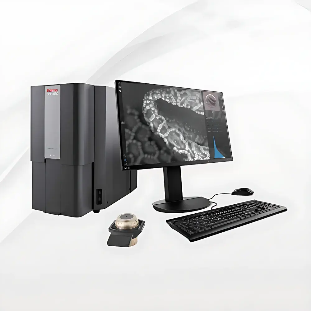
Phenom ProX_1 Desktop Scanning Electron Microscope for Metallographic Analysis
| Brand | Phenom |
|---|---|
| Origin | Netherlands |
| Model | Phenom ProX_1 |
| Instrument Type | Benchtop SEM |
| Electron Source | Cerium Hexaboride (CeB₆) |
| Secondary Electron Resolution | 6 nm |
| Backscattered Electron Resolution | 8 nm |
| Magnification Range | Up to 350,000× |
| Accelerating Voltage | 4.8–20.5 kV |
Overview
The Phenom ProX_1 Desktop Scanning Electron Microscope is an engineered solution for routine and advanced metallographic characterization in industrial quality control, failure analysis, and materials R&D laboratories. Operating on the principle of scanning electron microscopy—where a focused beam of high-energy electrons interacts with conductive or coated metallic specimens to generate secondary electrons (SE), backscattered electrons (BSE), and characteristic X-rays—the ProX_1 delivers high-fidelity imaging and integrated energy-dispersive X-ray spectroscopy (EDS) capability in a compact, vibration-insensitive benchtop platform. Designed specifically for metallurgical applications, it enables direct correlation between microstructural morphology (e.g., grain boundaries, precipitates, fracture surfaces, phase distribution) and elemental composition without requiring ultra-high vacuum or complex sample preparation protocols typical of floor-standing systems. Its CeB₆ thermionic electron source ensures stable beam current and superior signal-to-noise ratio at low accelerating voltages—critical for minimizing charging artifacts on polished metal sections and preserving fine surface topography.
Key Features
- Benchtop architecture with integrated vacuum system: eliminates need for external pumps or dedicated lab infrastructure; achieves operational vacuum in under 30 seconds.
- Cerium hexaboride (CeB₆) electron gun: offers 5–10× longer lifetime and higher brightness than tungsten filaments, enabling consistent resolution down to 6 nm (SE) and 8 nm (BSE) across the full voltage range (4.8–20.5 kV).
- Automated Eucentric stage with 5-axis motorized control: supports precise tilt (±65°), rotation, and Z-height adjustment for comprehensive fracture surface inspection and cross-sectional imaging.
- Real-time drift compensation algorithm: maintains positional stability during long-duration acquisitions, essential for EDS mapping of alloy phases and inclusion populations.
- Integrated EDS detector with silicon drift detector (SDD) technology: enables rapid qualitative and semi-quantitative elemental analysis (Z ≥ 5) with detection limits <0.1 wt% for major alloying elements (Fe, Cr, Ni, Al, Ti, Mo).
- One-click automated workflow: includes SmartSEM software for guided alignment, auto-focus, auto-stigmation, and batch image acquisition—reducing operator dependency and enhancing inter-lab reproducibility.
Sample Compatibility & Compliance
The Phenom ProX_1 accommodates standard metallographic specimens up to 100 mm in diameter and 40 mm in height—including mounted and polished cross-sections, fractured tensile bars, fatigue crack tips, and sputter-coated powder compacts. Conductive coatings (e.g., carbon or gold/palladium) are optional for non-conductive oxide layers or ceramic inclusions. The system complies with IEC 61000-6-3 (EMC emissions) and IEC 61000-6-2 (immunity), and its software architecture supports audit trail functionality aligned with GLP and ISO/IEC 17025 requirements. While not certified to FDA 21 CFR Part 11 out-of-the-box, the SmartSEM platform allows configuration of user access levels, electronic signatures, and secure data export—facilitating integration into regulated environments where traceability of microstructural evidence is required (e.g., aerospace component certification per AMS 2640 or ASTM E3, E407).
Software & Data Management
SmartSEM v3.x provides a unified interface for instrument control, image acquisition, EDS spectral processing, and report generation. All acquired images and spectra are stored in vendor-neutral TIFF and .eds formats, with metadata embedded per EXIF standards. Batch processing tools support automated particle analysis (size, shape, circularity), phase segmentation via BSE intensity thresholding, and inclusion classification based on elemental ratios (e.g., Al₂O₃ vs. MnS vs. TiN). Data exports comply with ASTM E1351 for metallographic reporting and can be imported directly into third-party platforms such as Thermo Scientific Pathfinder or Bruker ESPRIT for advanced multivariate statistical analysis. Raw EDS spectra are compatible with NIST DTSA-II reference libraries for standardized peak deconvolution.
Applications
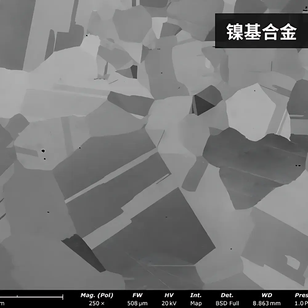
- Metal fracture surface analysis: identification of ductile/brittle transition zones, cleavage facets, and intergranular cracking mechanisms in steels, aluminum alloys, and nickel-based superalloys.
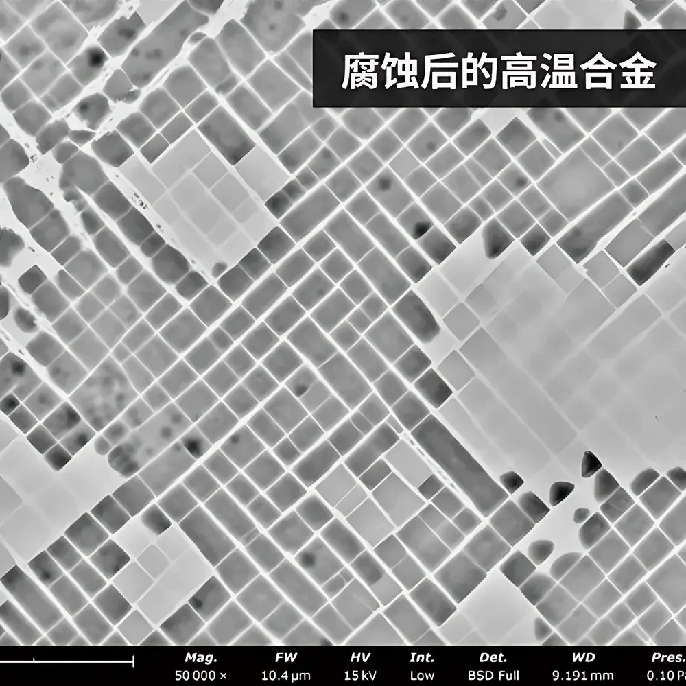
- Phase identification and distribution mapping: quantification of ferrite/austenite ratios in duplex stainless steels; carbide/nitride precipitation kinetics in heat-treated tool steels.
- Inclusion characterization: morphological classification and compositional fingerprinting of non-metallic inclusions (e.g., oxides, sulfides, silicates) per ASTM E45 and ISO 4967 protocols.
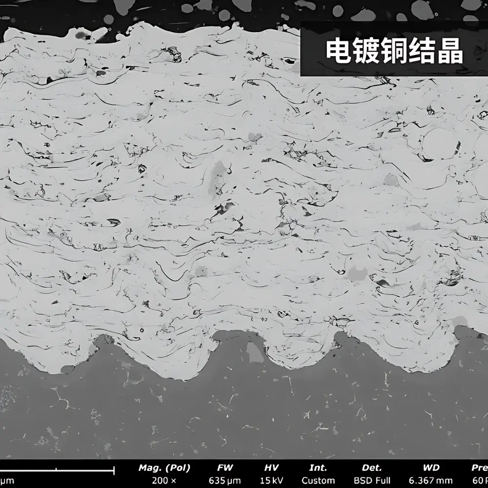
- Coating and interface analysis: thickness measurement of PVD/CVD layers, adhesion assessment at metal-ceramic interfaces, and oxidation scale growth evaluation after thermal cycling.
- Powder metallurgy QA: particle size distribution, sphericity assessment, and surface defect detection in gas-atomized Ni-based or Ti-6Al-4V powders prior to additive manufacturing.
FAQ
What sample preparation is required for metallic specimens?
Standard metallographic preparation—grinding, polishing, and optional etching—is sufficient. For non-conductive oxide scales or ceramic inclusions, a 5–10 nm carbon coat is recommended to prevent charging.
Can the Phenom ProX_1 perform quantitative EDS analysis?
Yes—it supports semi-quantitative analysis using ZAF matrix correction algorithms. For certified quantitative results (e.g., ASTM E1508), standards-based calibration with certified reference materials is advised.
Is the system compatible with ion milling or FIB-prepared cross-sections?
Yes. The large chamber and motorized stage accommodate ion-milled lamellae mounted on standard SEM stubs; tilt capability enables optimal viewing of subsurface features.
How does the CeB₆ source compare to field emission guns (FEG) in resolution performance?
While FEG-SEMs achieve sub-nm resolution under ideal conditions, the CeB₆ source delivers robust 6 nm SE resolution with superior stability, lower operating cost, and no requirement for ultra-high vacuum—making it optimal for routine metallurgical QA/QC.
Does the system support automated inclusion analysis per ASTM E45?
Yes. When paired with optional ParticleX Metal module, it executes fully automated detection, classification, and statistical reporting compliant with ASTM E45 Type A–D rating methodologies.


