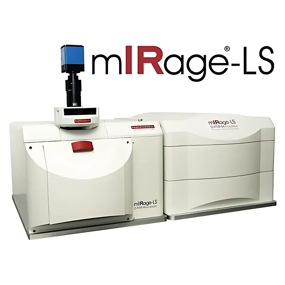
PSC mIRage-LS Optical Photothermal Infrared (O-PTIR) Microscope with Fluorescence and Raman Integration
| Brand | PSC |
|---|---|
| Origin | USA |
| Manufacturer Type | Authorized Distributor |
| Import Status | Imported |
| Model | mIRage-LS |
| Instrument Category | Laser-Based Infrared Spectrometer |
| Instrument Type | Laboratory Benchtop System |
| Wavenumber Range | 3600–800 cm⁻¹ |
| Spatial Resolution | ~500 nm |
Overview
The PSC mIRage-LS is a fully integrated optical photothermal infrared (O-PTIR) microscope engineered for label-free, sub-diffraction vibrational imaging of live biological specimens at unprecedented spatial fidelity. Unlike conventional Fourier-transform infrared (FTIR) microscopes constrained by the diffraction limit (~3–10 µm in mid-IR), the mIRage-LS employs pulsed tunable quantum cascade laser (QCL) excitation coupled with synchronized visible probe beam detection to achieve photothermal expansion-induced signal generation—bypassing Mie scattering artifacts and enabling true <500 nm lateral resolution in both reflection and transmission geometries. This non-contact, far-field technique delivers chemically specific IR absorption contrast without requiring attenuated total reflection (ATR) or vacuum environments, making it uniquely suited for hydrated, unstained, and dynamic biological systems—including live mammalian cells in physiological buffer, intact tissue sections, and single bacterial or viral particles. The system’s core architecture supports concurrent acquisition of IR, Raman, and fluorescence signals from identical subcellular coordinates, establishing a rigorous platform for multimodal correlative spectroscopy under native conditions.
Key Features
- Simultaneous O-PTIR, confocal Raman, and widefield fluorescence imaging on a single optical platform
- Sub-500 nm spatial resolution across full 3600–800 cm⁻¹ spectral range—enabling direct mapping of protein secondary structure, lipid acyl chains (e.g., CH₂/CH₃ at 2856/2874 cm⁻¹), amide I/II, and nucleic acid vibrations at biologically relevant length scales
- True non-contact measurement: no physical contact with sample surface; compatible with aqueous media, soft tissues, and delicate monolayers
- Dual-mode operation: reflection geometry optimized for thick or uneven samples (e.g., tissue sections, biofilms); transmission mode enabled for thin films and cultured cells
- Integrated dual-tuned QCL source covering both C–H stretching (3000–2700 cm⁻¹) and fingerprint regions (1800–950 cm⁻¹), ensuring comprehensive molecular coverage without mechanical realignment
- Real-time spectral acquisition at pixel dwell times as low as 10 ms, supporting time-resolved studies of cellular dynamics (e.g., drug uptake, lipid droplet trafficking)
Sample Compatibility & Compliance
The mIRage-LS accommodates native-state biological specimens without fixation, dehydration, or metal coating—preserving structural integrity and biochemical functionality. Validated use cases include live adherent and suspension cells (e.g., MIA PaCa-2, erythrocytes), cryo-sectioned and FFPE tissue slices (up to 10 µm thickness), bacterial colonies, isolated virions, and extracellular vesicles. Its open optical path permits integration with environmental chambers (temperature, CO₂, humidity control) and microfluidic devices for long-term live-cell experiments. From a regulatory standpoint, spectral data acquisition complies with GLP-aligned metadata logging (timestamp, laser power, detector gain, stage position). While not inherently 21 CFR Part 11-compliant, the system supports audit-trail-ready software configurations when deployed with validated LIMS interfaces and electronic signature modules per institutional SOPs.
Software & Data Management
Acquisition and analysis are managed via PSC’s proprietary mIRage Suite v4.x, a modular application built on a Python-based computational framework. Core capabilities include hyperspectral cube generation (wavenumber × x × y), multivariate curve resolution (MCR), principal component analysis (PCA), hierarchical clustering, and co-localization quantification (Pearson/Spearman correlation between IR/Raman/fluorescence channels). Spectral libraries (Wiley KnowItAll, BioRad, custom-built) support automated peak matching with >95% confidence scoring against reference spectra. All raw interferograms, photothermal transients, and processed maps are stored in HDF5 format with embedded EXIF-style metadata—ensuring FAIR (Findable, Accessible, Interoperable, Reusable) data principles. Batch processing pipelines enable reproducible preprocessing (baseline correction, vector normalization, SNV) across multi-user laboratories.
Applications
- Single-cell phenotyping: Discrimination of normal vs. diseased cell states via IR-defined lipid-to-protein ratios, glycogen accumulation, or oxidative stress markers (e.g., carbonyl stretch at 1720 cm⁻¹)
- Drug–cell interaction kinetics: Time-lapse tracking of small-molecule diffusion, intracellular binding, and metabolic perturbation using marker bands (e.g., C=O ester hydrolysis at 1735 cm⁻¹)
- Lipid droplet biophysics: Sub-200 nm mapping of saturated vs. unsaturated acyl chain order in live adipocytes or hepatocytes—resolving heterogeneity masked by water absorption in conventional FTIR
- Neurodegenerative pathology: Co-localized detection of β-sheet amyloid aggregates (1620 cm⁻¹) and redox-active iron clusters (via S-XRF correlation) in Alzheimer’s tissue sections
- Microbial identification: Strain-level differentiation of Gram-positive/negative bacteria based on peptidoglycan (1450 cm⁻¹) and LPS (1240 cm⁻¹) spectral signatures at single-cell resolution
- Virology: Structural characterization of individual virions (e.g., vaccinia virus) including capsid protein conformation and envelope lipid composition
FAQ
How does O-PTIR overcome the diffraction limit in infrared microscopy?
O-PTIR uses a visible probe beam to detect nanoscale thermal expansion induced by pulsed mid-IR absorption—decoupling spatial resolution from IR wavelength. This photothermal mechanism achieves ~500 nm resolution independent of the 3–10 µm diffraction barrier inherent to direct IR detection.
Can the mIRage-LS acquire data from live cells in aqueous medium?
Yes. The non-contact, reflection-mode O-PTIR configuration eliminates water absorption artifacts that dominate transmission FTIR. Live cells in PBS or culture medium can be imaged continuously for >60 minutes without photodamage or thermal drift.
What spectral databases are supported for automated biomolecular assignment?
The system integrates Wiley KnowItAll, BioRad’s IR Biomaterials Library, and user-curated collections. Match confidence scores ≥95% are routinely achieved for proteins, lipids, carbohydrates, and nucleic acids in biological matrices.
Is polarization control available for anisotropy studies?
Yes. An optional motorized wire-grid polarizer enables controlled IR beam polarization—critical for collagen fiber orientation mapping and membrane lipid order assessment.
How is data interoperability ensured across multi-instrument workflows?
HDF5-formatted datasets include standardized metadata headers compliant with NIH-supported BIDS-IR extensions, enabling seamless import into MATLAB, Python (scikit-learn, HyperSpy), and commercial platforms like Thermo Scientific OMNIC.