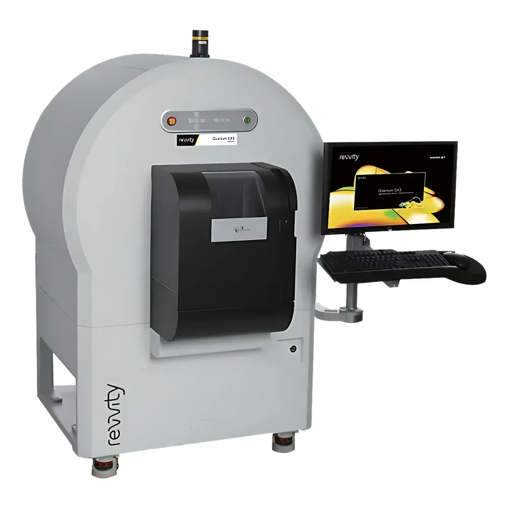
Revvity Quantum GX3 microCT Small Animal In Vivo Imaging System
| Brand | Revvity |
|---|---|
| Origin | USA |
| Manufacturer Type | Original Equipment Manufacturer (OEM) |
| Import Status | Imported |
| Model | Quantum GX3 |
| Instrument Type | X-ray Computed Tomography (microCT) |
| Animal Models Supported | Mice, Rats, Guinea Pigs, Rabbits, Zebrafish, Insects, Excised Tissues & Organs |
| Spatial Resolution | ≤5 µm |
| Voxel Size | Down to 2.3 µm |
| Scan Time (Minimum) | 3.9 s |
| Field of View (FOV) | 8, 18, 36, 72, and 86 mm |
| Filter Options | Six motorized, auto-sensed filters — Al (0.5 mm, 1.0 mm), Cu (0.1 mm, 0.2 mm, 1.0 mm), Al 0.5 mm + Cu 0.06 mm |
| Gating Capabilities | Dual-phase retrospective respiratory and cardiac gating |
| Modality Integration | Native co-registration with IVIS Spectrum optical imaging systems |
Overview
The Revvity Quantum GX3 microCT Small Animal In Vivo Imaging System is a high-performance, benchtop X-ray computed tomography platform engineered for non-invasive, quantitative 3D structural imaging of live small animals and excised biological specimens. Operating on the principle of cone-beam microCT, the system employs a high-stability microfocus X-ray source and a large-area flat-panel detector to acquire projection data from multiple angular positions. Reconstruction algorithms—based on filtered back-projection and iterative techniques—generate isotropic volumetric datasets with sub-5 µm spatial resolution. Designed specifically for longitudinal preclinical research, the Quantum GX3 delivers consistent image quality across repeated in vivo scans while maintaining radiation doses well within ALARA (As Low As Reasonably Achievable) guidelines for murine and lagomorph models. Its modular architecture supports both rapid whole-body surveys and high-resolution regional acquisitions—enabling morphometric, densitometric, and dynamic functional assessments without tissue disruption or contrast agent dependency.
Key Features
- Sub-5 µm spatial resolution with voxel sizes down to 2.3 µm—optimized for trabecular bone microarchitecture, alveolar septal thickness, and microvascular calcification analysis.
- Ultra-fast acquisition: Full-volume scans completed in as little as 3.9 seconds, minimizing motion artifacts and enabling high-throughput screening protocols.
- Five selectable field-of-view configurations (8–86 mm diameter) accommodate diverse sample geometries—from zebrafish embryos and insect exoskeletons to intact rabbit thoraces—without hardware reconfiguration.
- Motorized, auto-sensed filter wheel with six attenuation options (Al, Cu, and hybrid combinations) ensures optimal beam hardening correction and contrast-to-noise ratio across tissue densities.
- Dual-phase retrospective gating synchronizes data acquisition with both respiratory and cardiac cycles, permitting motion-compensated imaging of lungs, heart chambers, and perfused vasculature.
- Integrated dose monitoring and real-time exposure feedback comply with institutional radiation safety policies and support IACUC-mandated dose reporting.
Sample Compatibility & Compliance
The Quantum GX3 accommodates a broad spectrum of biological specimens: live anesthetized rodents (mice, rats, guinea pigs, rabbits), aquatic models (zebrafish larvae), arthropods (Drosophila, cockroaches), and ex vivo tissues (femoral heads, lung lobes, tumor xenografts, decalcified bone sections). All scanning protocols adhere to widely accepted preclinical imaging standards, including ASTM E1441 (Standard Practice for Computed Tomography), ISO 15739 (Noise Measurement in Digital Radiography), and NIH/NCI guidelines for longitudinal microCT study design. The system’s software enforces audit-trail logging per GLP requirements and supports 21 CFR Part 11-compliant user authentication and electronic signature workflows when deployed in regulated environments.
Software & Data Management
Acquisition, reconstruction, and quantification are unified under the Quantum GX3’s proprietary software suite—compatible with Windows 10/11 64-bit platforms. Reconstruction engines support GPU-accelerated iterative algorithms (e.g., SART, OS-SART) and provide optional beam-hardening correction, ring artifact suppression, and phase-retrieval enhancements. Quantitative modules include BoneJ (for trabecular thickness, spacing, and connectivity density), LungQuant (for airway lumen area, parenchymal density distribution), and VesselQuant (for vessel volume, branching angle, and tortuosity metrics). DICOM 3.0 export enables PACS integration, while NIfTI and TIFF stack outputs facilitate import into MATLAB, Amira, Avizo, or 3D Slicer for advanced segmentation and mesh generation. Raw projection data and metadata (including kVp, mAs, filter ID, gating timestamps) are archived in vendor-neutral formats compliant with FAIR data principles.
Applications
- Bone & Mineral Research: Longitudinal assessment of osteoporosis progression, fracture healing, biomaterial integration, and metastatic bone lesion burden using BMD (bone mineral density) and microarchitectural parameters.
- Pulmonary Imaging: Quantification of emphysema severity, fibrosis extent, airway remodeling, and ventilation heterogeneity in murine asthma, COPD, and IPF models.
- Cardiovascular Phenotyping: Measurement of left ventricular mass, ejection fraction, wall thickness dynamics, and coronary calcification volume in hypertensive or atherosclerotic models.
- Oncology: Volumetric tracking of orthotopic or subcutaneous tumor growth, necrotic core development, and vascular co-option via contrast-enhanced CT angiography.
- Multimodal Correlative Studies: Precise spatial registration of bioluminescent/optical signals (from IVIS Spectrum) onto anatomical CT volumes enables cell trafficking, reporter gene expression, and therapy response mapping.
FAQ
What animal species are validated for use with the Quantum GX3?
Mice, rats, guinea pigs, rabbits, zebrafish, and insects are routinely imaged; protocols for larger species (e.g., ferrets, mini-pigs) may be developed subject to FOV and dose constraints.
Does the system support contrast-enhanced CT imaging?
Yes—iodinated or nanoparticle-based contrast agents can be administered intravenously or via inhalation; software includes dedicated enhancement subtraction and kinetic modeling tools.
Can raw projection data be exported for third-party reconstruction?
Yes—unprocessed sinograms and metadata are accessible via secure file export in HDF5 format, supporting custom algorithm development and validation.
Is respiratory and cardiac gating performed prospectively or retrospectively?
Both modes are available; retrospective dual-gating is standard, allowing flexible post-hoc sorting of projections based on recorded physiological waveforms.
How does the Quantum GX3 ensure compliance with institutional radiation safety programs?
The system logs all exposure parameters, provides real-time dose estimation (in mGy), and integrates with facility-wide dosimetry management systems via HL7 or CSV export.


