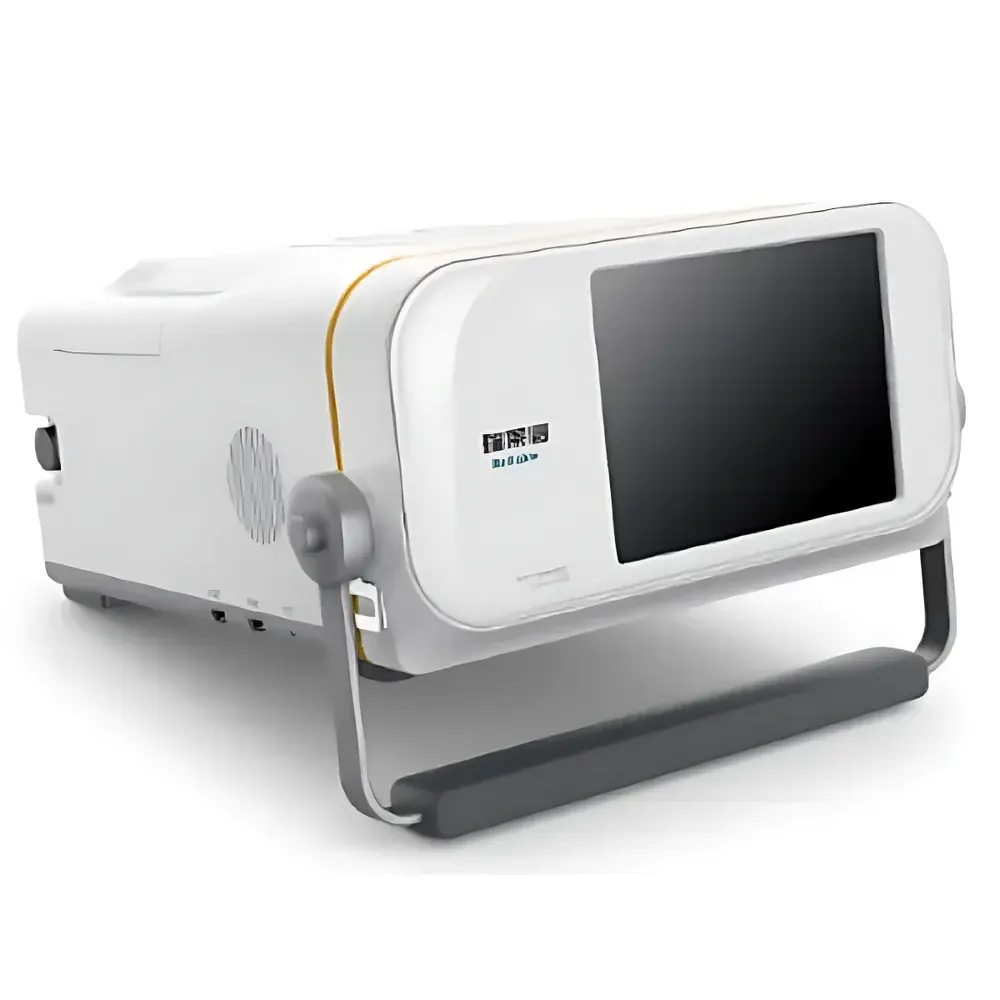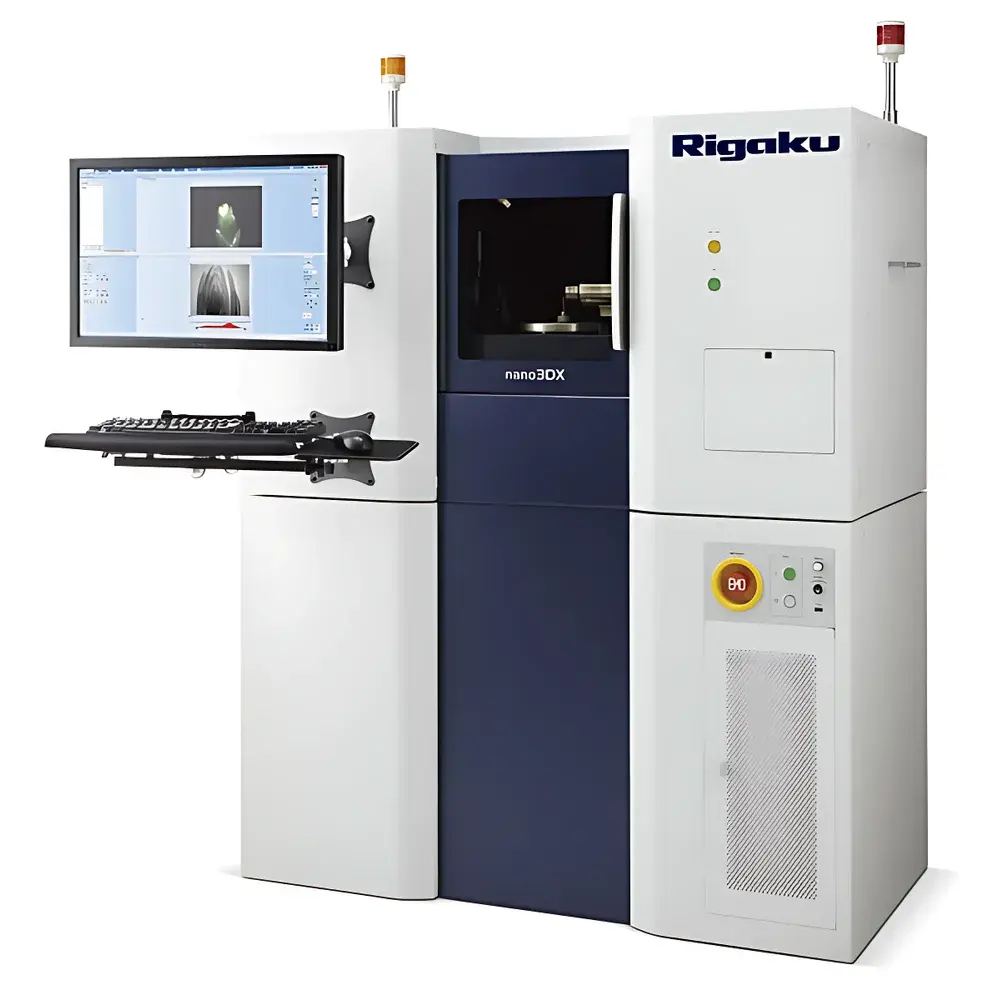
Rigaku Nano3DX and 3D CT Systems – High-Resolution X-Ray Microtomography Platforms
| Brand | Rigaku |
|---|---|
| Origin | Japan |
| Model | Nano3DX & 3D CT |
| Configuration | Benchtop and Floor-Standing Options |
| Application Scope | Universal Materials Characterization |
| Elemental Analysis Capability | Not Applicable (Non-destructive Imaging, Not Spectrometric) |
| Spatial Resolution | Sub-micron (Nano3DX), Micron-scale (3D CT) |
| Field of View | Adjustable from ~100 µm to >50 mm |
| Contrast Sensitivity | Optimized for Density Variations in Low-Z and Medium-Z Materials |
| Repeatability | Engineered for high scan-to-scan reproducibility under controlled environmental conditions |
| Compliance | Designed to support GLP/GMP-aligned workflows |
Overview
The Rigaku Nano3DX and 3D CT systems are benchtop and floor-standing X-ray microtomography (micro-CT) platforms engineered for non-destructive, high-fidelity 3D internal structure visualization across diverse solid-state materials. Unlike energy-dispersive X-ray fluorescence (EDXRF) spectrometers—which perform elemental quantification via characteristic X-ray emission—the Nano3DX and 3D CT operate on the principle of absorption-based X-ray computed tomography. They reconstruct volumetric datasets by acquiring hundreds to thousands of 2D projection images as the sample rotates through 360°, followed by filtered back-projection or iterative reconstruction algorithms. The Nano3DX leverages Rigaku’s proprietary high-brightness rotating anode X-ray source and a high-dynamic-range, low-noise CCD-based detector to achieve true sub-micron isotropic voxel resolution (down to 0.3 µm), enabling quantitative morphological analysis of fine microstructures such as nanopores, grain boundaries, fiber networks, and interfacial phases. The broader 3D CT platform offers scalable resolution and field-of-view configurations—from micron-level detail in small specimens to centimeter-scale volumes—without requiring sectioning, coating, or vacuum-compatible preparation.
Key Features
- Sub-micron spatial resolution (Nano3DX) and scalable micron-to-millimeter resolution (3D CT), supporting multi-scale structural interrogation
- High-contrast density discrimination enabled by optimized polychromatic X-ray spectrum and high quantum efficiency detection
- Full 360° continuous rotation stage with sub-arcsecond angular precision and motorized sample positioning
- Integrated thermal management and vibration-damped mechanical architecture for long-duration, high-stability scans
- Benchtop (Nano3DX) and floor-standing (3D CT) configurations to accommodate lab space constraints and large-sample throughput needs
- Automated alignment routines, beam hardening correction, ring artifact suppression, and phase-retrieval-ready acquisition modes
Sample Compatibility & Compliance
The systems accept a wide range of solid, non-liquid samples—including metals, ceramics, polymers, composites, biological tissues (fixed or dehydrated), geological specimens, electronic assemblies, and additively manufactured parts—without destructive sectioning or conductive coating. Maximum sample dimensions supported range from <1 mm³ (for nano-resolution imaging) up to 150 mm diameter × 200 mm height (3D CT). Both platforms comply with IEC 61000-6-3 (EMC emissions) and IEC 61000-6-2 (immunity) standards. While not medical devices, they support research-grade imaging workflows aligned with ISO 16234 (microstructural characterization of porous materials), ASTM E1441 (standard guide for computed tomography), and USP (imaging methods for pharmaceutical solid dosage forms). Audit-trail-capable acquisition logs and metadata embedding support GLP/GMP documentation requirements.
Software & Data Management
Acquisition and reconstruction are managed via Rigaku’s proprietary CT-SOLUTION software suite, featuring intuitive workflow-driven interfaces for scan planning, real-time projection monitoring, and GPU-accelerated 3D reconstruction (FDK, SART, and iterative regularization options). Post-processing tools include segmentation (threshold, region-growing, machine-learning-assisted), pore network extraction, skeletonization, thickness mapping, and quantitative morphometry (porosity %, specific surface area, tortuosity, connectivity density). Export formats include DICOM, TIFF stacks, STL, OBJ, and HDF5—ensuring compatibility with third-party analysis platforms (Avizo, Dragonfly, ImageJ/Fiji, MATLAB). All processing steps are timestamped and parameter-logged, satisfying traceability requirements for regulated environments.
Applications
These systems serve as core infrastructure for advanced materials R&D and quality assurance across sectors including battery electrode architecture analysis (Li-ion anode/cathode porosity gradients), additive manufacturing defect detection (lack-of-fusion, residual stress-induced cracking), polymer composite fiber orientation quantification, pharmaceutical tablet coating uniformity assessment, semiconductor package void analysis, aerospace component integrity verification, paleontological fossil digitization, and forensic fracture surface reconstruction. Time-resolved (4D) scanning—enabled by repeatable mounting and thermal/environmental control—is routinely applied to in situ aging, thermal expansion, hydration/dehydration, and mechanical loading studies.
FAQ
Do the Nano3DX and 3D CT systems perform elemental analysis?
No. These are X-ray microtomography instruments designed exclusively for 3D density-based structural imaging—not elemental spectroscopy. For elemental composition, Rigaku offers separate EDXRF (e.g., NEX CG series) or WDXRF platforms.
Can these systems image hydrated or soft biological tissues?
Yes—with appropriate fixation (e.g., glutaraldehyde/osmium tetroxide) or cryo-fixation. Live or unfixed aqueous samples are not compatible due to radiation damage and lack of structural stability under vacuum or air-scanning conditions.
What is the typical scan time for a full 3D reconstruction?
Scan duration depends on resolution, sample size, and signal-to-noise requirements—ranging from 15 minutes (low-res, large FOV) to >24 hours (sub-micron Nano3DX acquisitions with high frame averaging).
Is remote operation and data transfer supported?
Yes. CT-SOLUTION supports secure remote desktop access, automated data export to network storage, and integration with LIMS via configurable API hooks for metadata synchronization.