RITE XSight™ FC Fiber-Optic Coupled High-Sensitivity X-ray Camera
| Brand | RITE |
|---|---|
| Origin | Czech Republic |
| Model | XSight™ FC |
| Sensor Types | Full-Frame CCD (FC5400, FC2160), sCMOS (μRapid) |
| Effective Pixels | 3326 × 2504 (CCD), 2048 × 2048 (sCMOS) |
| Field of View | 18.0 mm × 13.5 mm (FC5400), 7.2 mm × 5.4 mm (FC2160), 13.3 mm × 13.3 mm (μRapid) |
| Measured Spatial Resolution | 16 µm @ 8 keV (FC5400), 8 µm @ 8 keV (FC2160), 20 µm @ 8 keV (μRapid) |
| X-ray Energy Range | 5–30 keV |
| Read Noise | 10 e⁻ RMS (FC5400), 7.5 e⁻ RMS (FC2160), 1.5 e⁻ RMS (fast scan) / 1.4 e⁻ RMS (slow scan) (μRapid) |
| Dark Current | 0.02 e⁻/pix/s @ −30 °C (CCD), 0.5 e⁻/pix/s @ +5 °C (μRapid) |
| Frame Rate | 1 fps (CCD), up to 100 fps (μRapid, fast scan), 35 fps (μRapid, slow scan) |
| Dynamic Range | 3100:1 (70 dB, CCD), 20000:1 (fast scan) / 21430:1 (slow scan) (μRapid) |
| Interface | USB 3.0 (CCD), Camera Link Full (sCMOS) |
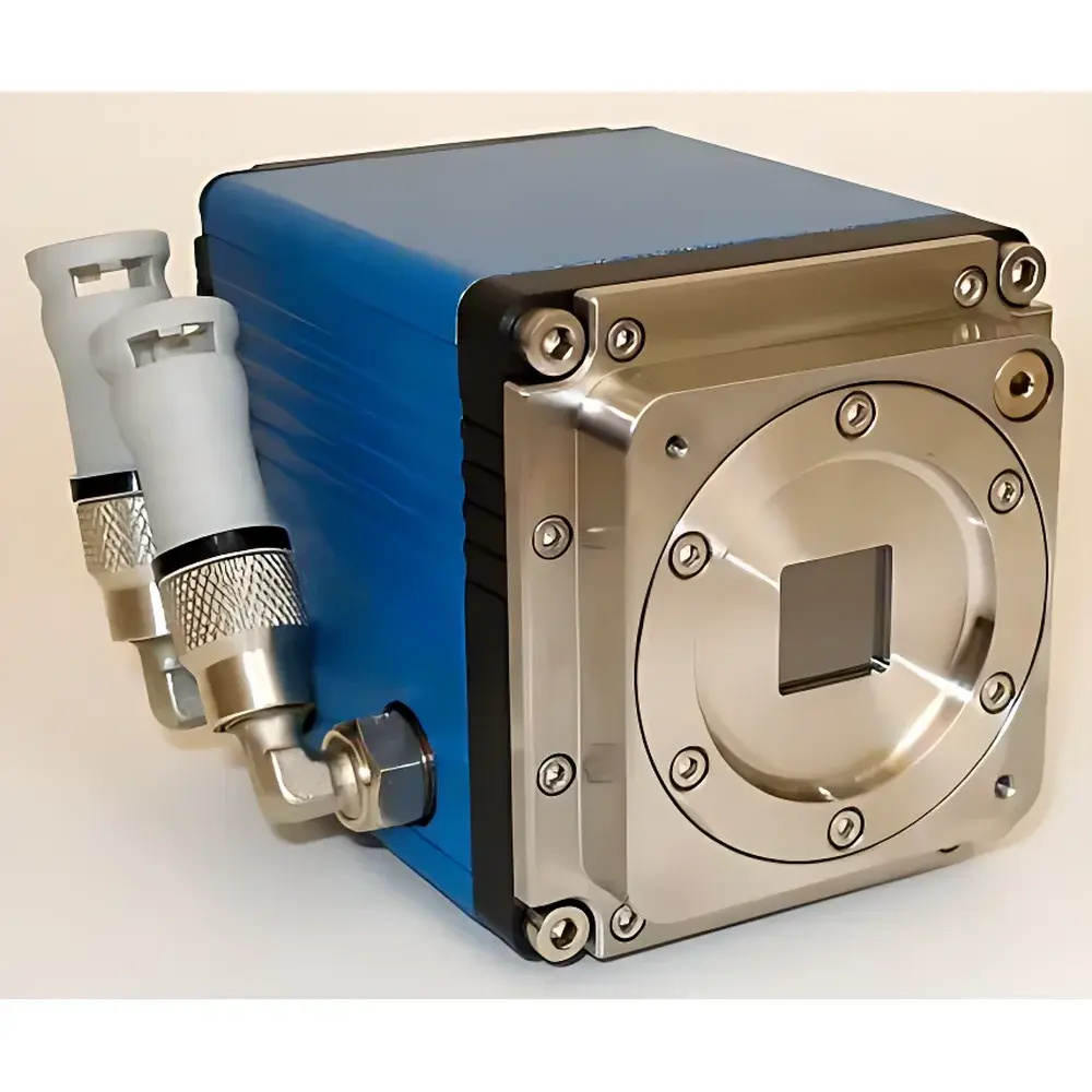
| Mounting Thread | M6 (CCD), ¼"-20 UNC (sCMOS) |
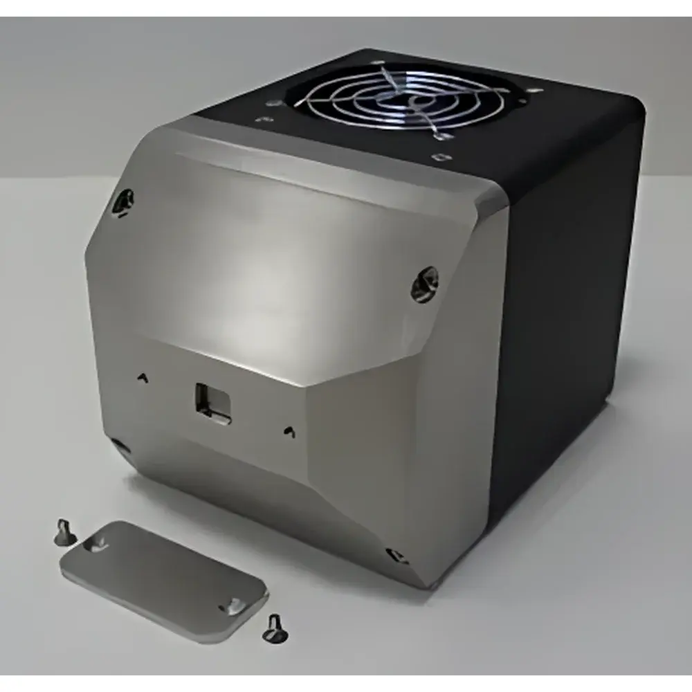
| Cooling | Thermoelectric (−30 °C for CCD |
| Optical Coupling | Tapered fiber-optic faceplate (no lens coupling) |
Overview
The RITE XSight™ FC is a high-performance, fiber-optic coupled X-ray imaging camera engineered for demanding synchrotron, laboratory-based X-ray microscopy, topography, and metrology applications. Unlike lens-coupled detectors, the XSight™ FC integrates a thin scintillator screen directly bonded to a tapered fiber-optic conduit—enabling photon-limited detection with ~20× higher quantum efficiency at typical hard X-ray energies (5–30 keV). This architecture preserves spatial fidelity while maximizing signal-to-noise ratio (SNR), particularly critical for low-dose or time-resolved measurements. The system comprises three variants: the XSight™ FC5400 and FC2160 full-frame CCD models optimized for high dynamic range and ultra-low noise in static or quasi-static imaging, and the XSight™ μRapid sCMOS variant designed for high-speed acquisition with sub-electron read noise and dual-scan modes. All models operate without water cooling—relying on precisely stabilized thermoelectric cooling—and feature compact, radiation-hardened mechanical housings that shield the sensor from stray X-ray scatter, thereby minimizing background-induced pixel corruption.
Key Features
- Fiber-optic coupling architecture delivering ~20× higher sensitivity vs. lens-coupled alternatives at 8 keV, validated by measured modulation transfer function (MTF) and detective quantum efficiency (DQE) curves.
- Three sensor configurations: FC5400 (3326 × 2504, 18 mm × 13.5 mm FOV), FC2160 (3326 × 2504, 7.2 mm × 5.4 mm FOV), and μRapid (2048 × 2048, 13.3 mm × 13.3 mm FOV), enabling scalable resolution–field trade-offs.
- Thermoelectric cooling to −30 °C (CCD) or +5 °C (sCMOS), eliminating dependency on external chillers and ensuring long-term thermal stability under vacuum or inert-gas environments.
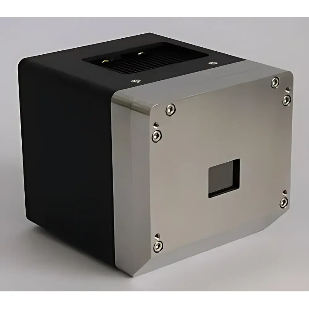
- Radiation-shielded aluminum housing with integrated M6 (CCD) or ¼”-20 UNC (sCMOS) mounting threads for direct integration into goniometers, diffractometers, or custom X-ray beamlines.
- USB 3.0 (CCD) and Camera Link Full (sCMOS) interfaces compliant with GenICam standard, supporting hardware-triggered acquisition, ROI streaming, and precise exposure synchronization.
- No moving parts, no liquid coolant, and fully sealed optical path—designed for GLP-compliant operation in regulated analytical laboratories.
Sample Compatibility & Compliance
The XSight™ FC supports non-destructive imaging of crystalline and polycrystalline materials—including silicon wafers, III-V semiconductors, quartz substrates, and biological specimens—under both monochromatic and white-beam illumination. Its 5–30 keV energy response aligns with common laboratory X-ray sources (e.g., Cu Kα, Mo Kα) and synchrotron bending-magnet beamlines. The detector meets ISO 11146-2 (laser beam profiling) and ASTM E94/E1441 (radiographic imaging standards) requirements for spatial resolution reporting. For regulated environments, firmware supports audit-trail logging and user-access control per FDA 21 CFR Part 11 when used with validated acquisition software. Mechanical design conforms to IEC 61000-6-2 (EMC immunity) and IEC 61000-6-4 (EMC emissions) for integration into ISO Class 5 cleanroom-compatible setups.
Software & Data Management
Acquisition and analysis are supported via RITE’s XSight Control Suite—a cross-platform application (Windows/Linux) providing real-time histogram equalization, flat-field correction, dark-frame subtraction, and multi-frame averaging. Raw data export is available in HDF5, TIFF, and FITS formats, preserving bit-depth integrity for downstream processing in Python (NumPy/SciPy), MATLAB, or commercial packages such as ImageJ/Fiji and Avizo. The SDK includes C/C++ and Python APIs compliant with GenICam v3.1, enabling seamless integration into custom LabVIEW, EPICS, or Tango-based control systems. All metadata—including exposure time, temperature, gain, and timestamp (with microsecond precision)—is embedded in image headers for traceability in GxP workflows.
Applications
- X-ray topography: Defect mapping in single-crystal ingots (e.g., Si, GaAs) using Berg-Barrett or section topography geometries.
- X-ray microscopy: Sub-20 µm resolution imaging of microstructures in battery electrodes, catalyst layers, and additive-manufactured alloys.
- Beamline alignment & diagnostics: Real-time monitoring of X-ray optics performance (mirrors, multilayers, KB lenses) via wavefront sensing and intensity profiling.
- Time-resolved radiography: In situ deformation studies (e.g., tensile testing, thermal cycling) leveraging the μRapid’s 100 fps full-resolution capability.
- Phase-contrast imaging: Edge-enhancement visualization of low-Z materials (polymers, soft tissues) without contrast agents, enabled by high DQE at low fluence.
FAQ
What is the primary advantage of fiber-optic coupling over lens coupling in X-ray imaging?
Fiber-optic coupling eliminates air gaps and refractive losses inherent in lens-based systems, resulting in higher X-ray photon collection efficiency—particularly critical below 20 keV—and improved spatial resolution retention due to reduced light spread within the taper.
Can the XSight™ FC be operated in vacuum or helium-purged environments?
Yes—the sealed aluminum housing and absence of outgassing elastomers or adhesives permit operation at pressures down to 10⁻³ mbar; optional O-ring sealed feedthroughs are available for Camera Link and power connectors.
Is dark current compensation automated during acquisition?
Yes—real-time dark frame subtraction is implemented in firmware, with user-selectable reference frame acquisition intervals (from single-shot to periodic calibration every 10 minutes) to accommodate thermal drift.
Does the system support synchronized multi-camera acquisition?
Yes—via TTL trigger input/output ports and programmable delay generators, enabling precise temporal alignment across multiple XSight™ FC units or hybrid X-ray/optical setups.
Are calibration certificates provided with each unit?
Each camera ships with a factory-issued calibration report including measured MTF at 8 keV, DQE(0), dark current vs. temperature, and linearity verification per ISO 15739, traceable to PTB (Physikalisch-Technische Bundesanstalt) standards.


