Sigray AttoMap-310 Wavelength Dispersive X-Ray Fluorescence Microscope
| Brand | Sigray |
|---|---|
| Origin | Shanghai, China |
| Model | AttoMap-310 |
| Spatial Resolution (with high-res optics) | 3–5 µm |
| Detection Sensitivity | sub-ppm (relative), femtogram-level (absolute) |
| Incident Angle Range | 3° to 90° (0.01° step) |
| X-ray Source | Patented microfocus source with up to 5 selectable anode targets (e.g., Si, Cr, Cu, Rh, Mo, W, Au, Ti, Ag) |
| Max Power / Voltage | 50 W / 20–45 kVp |
| X-ray Optics | Dual-parabolic grazing-incidence mirrors, ~80% transmission efficiency, 1:1 default magnification |
| Detector | Silicon Drift Detector (SDD), energy resolution <129 eV (Mn-Kα) |
| Vacuum Chamber | <10⁻⁴ Torr |
| Sample Stage Travel | 100 × 100 mm |
| Light Element Detection Limit | Down to boron (B), quantitative for C, N, O at <1% wt. |
Overview
The Sigray AttoMap-310 is a laboratory-based wavelength dispersive X-ray fluorescence (WDXRF) microscope engineered for ultra-high spatial resolution elemental mapping and quantitative microanalysis. Unlike conventional energy-dispersive micro-XRF (ED-microXRF) systems—whose spatial resolution is typically limited to 20–50 µm—the AttoMap-310 achieves true microscale imaging down to 3–5 µm using proprietary dual-parabolic X-ray focusing optics. This capability is enabled by a patented high-brightness microfocus X-ray source coupled with precision grazing-incidence mirror optics, delivering >50× higher beam brightness than competing microXRF platforms. The system operates on fundamental XRF physics: incident X-rays eject inner-shell electrons from sample atoms; characteristic fluorescent X-rays emitted during electron relaxation are energy-resolved and spatially mapped to generate quantitative elemental distribution images. Its design supports both vacuum (<10⁻⁴ Torr) and ambient operation, making it uniquely suited for trace-level light-element analysis (Z ≥ 5, including B, C, N, O) as well as high-sensitivity heavy-element detection across semiconductor, geological, biological, and battery materials.
Key Features
- Sub-5 µm spatial resolution enabled by Sigray’s proprietary dual-parabolic X-ray focusing optics—eliminating chromatic aberration inherent in polycapillary lenses.
- Patented multi-target microfocus X-ray source supporting up to five selectable anode materials (e.g., Si, Cr, Cu, Rh, Mo, W, Au, Ti, Ag), enabling energy-tunable excitation optimized per element group.
- Motorized goniometric stage permitting precise incident angle control from 3° (grazing incidence) to 90° (normal incidence) in 0.01° increments—critical for thin-film analysis, diffraction suppression on crystalline substrates (e.g., Si wafers), and enhanced signal-to-background for low-Z elements.
- Ultra-high-vacuum chamber (<10⁻⁴ Torr) with platinum-coated internal surfaces, ensuring optimal transmission of low-energy X-rays (e.g., C Kα = 277 eV) and enabling quantitative organic matrix analysis at sub-1% wt. levels.
- High-efficiency silicon drift detector (SDD) with <129 eV Mn-Kα energy resolution and integrated optical microscope for correlative optical–XRF imaging.
- Modular software architecture supporting application-specific workflows—including semiconductor pattern recognition, AI-driven mineral clustering, and fundamental parameter (FP)-based quantification without standards.
Sample Compatibility & Compliance
The AttoMap-310 accommodates diverse sample geometries—from 300 mm semiconductor wafers and geological thin sections to cryo-preserved biological tissue slices and battery electrode cross-sections. Its variable-angle illumination and vacuum compatibility ensure robust performance across conductive, insulating, hydrated, and beam-sensitive specimens. For regulated environments, the system supports audit-ready data handling: raw spectra, acquisition metadata, and processing logs are stored in vendor-neutral HDF5 format with timestamped provenance. Software modules comply with GLP/GMP-aligned practices, including electronic signatures, version-controlled analysis protocols, and full audit trails—facilitating alignment with FDA 21 CFR Part 11 requirements where instrument qualification is required. While not certified to ISO/IEC 17025 out-of-the-box, the platform’s traceable calibration routines (using NIST-traceable reference materials) and documented uncertainty budgets enable laboratory-developed validation per ASTM E1621 (Standard Guide for XRF Elemental Analysis) and ISO 18507 (Micro-XRF for elemental mapping).
Software & Data Management
Sigray Composition™ serves as the unified control and analysis suite, featuring three interoperable modules: Semiconductor Acquisition (for recipe-driven wafer navigation and automated point analysis), GeoMineral (applying unsupervised machine learning to cluster spectral signatures and assign mineral phases), and LifeScience Quant (enabling FP-based quantification with or without standards). All modules support batch processing, spectral deconvolution, overlay of optical/XRF images, and export of quantitative maps in TIFF, CSV, or HDF5 formats. For advanced users, Python-accessible Jupyter Notebooks are provided—preloaded with API bindings to instrument control, spectral fitting libraries (e.g., PyMca), and FP modeling kernels—allowing custom algorithm development, statistical process control (SPC) integration, or ML model retraining. Data integrity is preserved via immutable project files containing full acquisition parameters, detector calibration history, and user-defined processing steps—ensuring reproducibility across laboratories and over time.
Applications
- Semiconductor Process Control: Quantitative mapping of dopants (e.g., B, P, As) and contaminants (e.g., Fe, Cu, Ni) in gate oxides and epitaxial layers; detection of sub-monolayer organic residues on 300 mm wafers under vacuum.
- Geosciences & Mineralogy: High-fidelity phase identification and trace-element zoning in ore minerals; simultaneous mapping of light elements (O, C, B) and transition metals (Fe, Cu, Zn) in petrographic thin sections.
- Life Sciences & Metallomics: Subcellular distribution of essential (Fe, Cu, Zn) and toxic (As, Cd) metals in frozen-hydrated tissue; longitudinal tracking of nanoparticle drug carriers in Daphnia models.
- Battery Materials: Localization of micron-scale metallic impurities (Cr, Hf, Zr) in cathode coatings; quantification of Li-loss gradients in NMC particles via indirect O/Ca ratio mapping.
- Environmental Botany: In planta mapping of hyperaccumulated metals (Ni, Zn, Se) in transgenic crops; correlation of nutrient uptake (K, Ca, Mg) with drought-stress biomarkers.
FAQ
What distinguishes the AttoMap-310 from standard ED-microXRF systems?
The AttoMap-310 employs wavelength-dispersive detection principles combined with microfocus excitation and dual-parabolic X-ray optics—achieving 3–5 µm resolution and sub-ppm sensitivity. In contrast, ED-microXRF relies on polycapillary optics and solid-state detectors with inherently lower peak-to-background ratios and poorer light-element response.
Can the system perform quantitative analysis without certified reference materials?
Yes. The fundamental parameter (FP) quantification engine uses first-principles physics—including X-ray mass attenuation coefficients, fluorescence yields, and detector response functions—to compute weight percentages directly from spectra, eliminating dependency on matrix-matched standards.
Is vacuum operation mandatory for light-element detection?
For quantitative analysis of elements below sodium (Z < 11), vacuum (<10⁻⁴ Torr) is required to prevent absorption of low-energy X-rays by air molecules. Ambient operation remains viable for heavier elements (e.g., Fe, Cu, Pb) where atmospheric attenuation is negligible.
How does the multi-target X-ray source improve analytical flexibility?
By selecting optimal anode materials (e.g., Cr for Ti/V detection, Mo for As/Se, Au for Pb/U), users maximize fluorescence cross-sections across the periodic table—improving signal intensity by up to three orders of magnitude versus fixed-anode sources.
Does the system support tomographic XRF (TXRF) or CL-XRFI acquisition?
Yes. The motorized goniometer and synchronized stage motion enable computerized laminography XRF imaging (CL-XRFI), allowing high-resolution 3D reconstruction of elemental distributions in thick or layered samples without destructive sectioning.
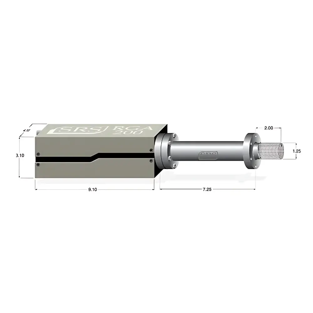
Related Products

")
")