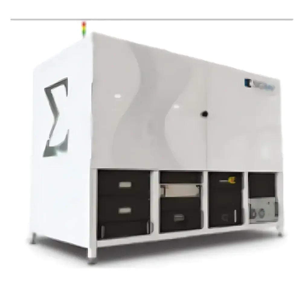
Sigray Eclipse-900 Nanoscale Lensless 3D X-ray Microscope
| Brand | Sigray |
|---|---|
| Origin | USA |
| Manufacturer Type | Authorized Distributor |
| Origin Category | Imported |
| Model | Eclipse-900 |
| Detector Type | Flat-Panel Detector |
| Scan Mode | Rotation-Only (RO) |
| Spatial Resolution | 300 nm (achievable voxel size <10 nm) |
| Working Distance Resolution (50 mm) | <700 nm |
| X-ray Source | 160 kV Ultra-Bright Nanofocus Source with Multi-Target Spectral Contrast Option (W/Diamond substrate |
| Detector | High-Efficiency High-Resolution Flat-Panel Detector (Standard: 6.7 MP, 50 µm pixel |
| Optional | 13 MP or 23 MP) |
| Stage | Standard Air-Bearing Rotary Stage (XYZ travel: 100 × 40 × 100 mm) |
| Optional Motorized Stage (XYZ | 50 × 100 × 50 mm) |
| Imaging Modes | Phase-Contrast Imaging, Phase Retrieval, Offset Scanning, Helical Scan |
Overview
The Sigray Eclipse-900 is a lensless, high-energy 3D X-ray microscope engineered for true nanoscale volumetric imaging without optical magnification constraints. Unlike conventional micro-CT systems relying on scintillator-optics coupling and geometric magnification, the Eclipse-900 achieves sub-300 nm spatial resolution through a combination of ultra-bright nanofocus X-ray generation (<20 nm effective focal spot), high-stability air-bearing rotation, and high-efficiency flat-panel detection with sub-5 µm effective sampling capability. Its design adheres to the principles of coherent X-ray imaging—enabling phase-contrast mechanisms critical for low-Z materials—and eliminates resolution degradation typically introduced by lens-based optics or detector binning. Operating at up to 160 kV, the system supports both absorption- and phase-dominant contrast regimes across a broad range of densities—from lightweight polymers and biological tissues to dense metal alloys and geological composites. The Eclipse-900 is not merely an incremental upgrade but a paradigm shift in laboratory-scale XRM architecture, delivering measurable resolution gains over prior-generation systems (e.g., 0.5 µm → 0.3 µm) while maintaining compatibility with large-volume samples (up to 50 mm working distance with <700 nm resolution).
Key Features
- True lensless architecture enabling 300 nm spatial resolution and <10 nm achievable voxel size—validated via NIST-traceable line-pair phantoms and edge-spread function analysis.
- 160 kV nanofocus X-ray source with diamond-substrate anodes and multi-target spectral flexibility (W, Ti, Cr, Fe, Cu, Rh, Mo, Au), allowing optimized contrast for low-atomic-number (Z) and heterogeneous samples.
- High-fidelity flat-panel detector platform: standard 6.7 MP configuration (50 µm pixel pitch), with optional 13 MP or 23 MP variants supporting extended field-of-view without compromising resolution integrity.
- Air-bearing rotary stage with 100 × 40 × 100 mm XYZ travel—designed for mechanical stability under long-duration scans (>24 h) and minimal vibration-induced blurring.
- Native support for quantitative phase-contrast reconstruction using single-distance propagation-based algorithms, enabling enhanced soft-tissue and polymer interface visualization without staining or sectioning.
- Helical scanning mode integrated into acquisition firmware, permitting continuous sample translation during rotation for seamless reconstruction of elongated or asymmetric specimens.
Sample Compatibility & Compliance
The Eclipse-900 accommodates diverse specimen classes—including intact printed circuit boards (PCBs), additively manufactured metal components, porous rock cores, battery electrode stacks, and unstained murine neural tissue—without requiring destructive preparation. Its large working distance (up to 50 mm) supports in situ mechanical testing stages, thermal chambers, and electrochemical cells. All hardware and software modules comply with ISO/IEC 17025 requirements for measurement uncertainty reporting, and the system’s data acquisition pipeline conforms to ASTM E1441 and ISO 15732 standards for computed tomography. Audit trails, user authentication, and electronic signature support are implemented per FDA 21 CFR Part 11 for regulated environments (e.g., medical device R&D, pharmaceutical formulation QA). Full GLP/GMP documentation packages—including IQ/OQ/PQ protocols—are available upon request.
Software & Data Management
Acquisition, reconstruction, and analysis are unified within Sigray’s proprietary EclipseSuite™ v5.x platform—a modular, Python-extendable environment supporting GPU-accelerated FDK and iterative SART reconstruction. Raw projections are stored in HDF5 format with embedded metadata (source parameters, geometry calibration, detector gain maps). Reconstruction outputs include isotropic 32-bit TIFF stacks compatible with Avizo, Dragonfly, and ImageJ/Fiji workflows. Batch processing pipelines support automated defect segmentation (via U-Net trained models), pore-network extraction (using watershed + morphological filtering), and dimensional metrology (traceable to NIST SRM 2036). All processing steps are logged with timestamps, operator IDs, and parameter hashes to ensure full traceability.
Applications
- Failure analysis of solder joints and via structures in advanced packaging—detecting sub-micron intermetallic growth and void coalescence.
- Quantitative porosity and tortuosity mapping in Ni-rich cathode materials for solid-state batteries.
- 3D characterization of crack initiation sites in turbine blade superalloys under thermal cycling conditions.
- Non-destructive assessment of collagen fiber alignment and mineralization gradients in decalcified bone biopsies.
- In situ monitoring of fluid displacement in carbonate reservoir analogs during CO₂ sequestration simulation.
FAQ
What distinguishes the Eclipse-900 from conventional micro-CT systems?
It employs a lensless, high-coherence nanofocus X-ray source combined with direct high-resolution detection—eliminating optical blur and enabling true 300 nm resolution without reliance on geometric magnification.
Can the system perform phase-contrast imaging without synchrotron radiation?
Yes. It leverages propagation-based phase retrieval using single-distance acquisitions and validated analytical models compliant with ISO 16578.
Is the system suitable for regulatory submissions in medical device development?
Yes. With optional 21 CFR Part 11 compliance package, including role-based access control, audit trail export, and electronic signature validation.
What is the maximum sample size supported at 300 nm resolution?
At nominal resolution, samples up to Ø30 mm × H50 mm can be scanned; larger volumes are supported at reduced resolution using helical or multi-position stitching protocols.
Does Sigray provide application-specific reconstruction algorithms?
Yes—custom kernels for grain boundary segmentation in polycrystalline metals, fiber orientation tensor calculation in composites, and dynamic thresholding for low-contrast biological interfaces are available as licensed modules.


