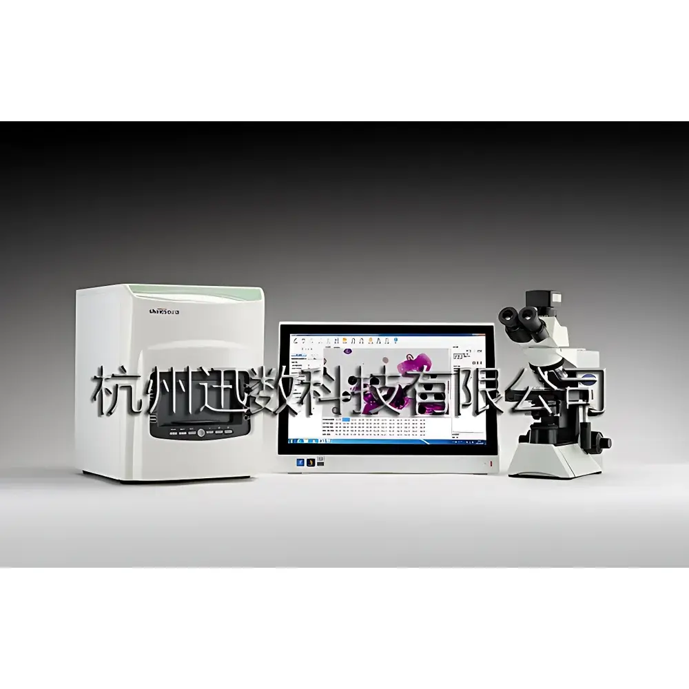
Xunshu GenTox 3 Integrated Micronucleus Analysis, Colony Counting & Cell Counting System
| Brand | Xunshu |
|---|---|
| Origin | Zhejiang, China |
| Manufacturer Type | OEM Manufacturer |
| Region Category | Domestic (China) |
| Model | GenTox 3 |
| Pricing | Upon Request |
Overview
The Xunshu GenTox 3 is an integrated digital image analysis platform engineered for standardized genetic toxicology assessment, microbial enumeration, and quantitative cell morphology analysis. It combines high-fidelity optical imaging with domain-specific AI-assisted algorithms to support regulatory-compliant studies under OECD 474, ISO 10993-3, ICH S2(R2), and USP / . The system centers on an Olympus CX31 upright microscope equipped with UIS2 infinity-corrected optics, a scientific-grade 2/3″ Sony ExView HAD CCD camera (ICX285AQ), and three tightly coupled software modules: Micronucleus Analysis Software (for polychromatic erythrocyte [PCE] identification and micronucleus scoring in bone marrow smears), MIC Cell Analysis Software (for morphometric quantification of stained cells or chromosomes), and Colony Analysis Software (for automated enumeration and classification of bacterial/fungal colonies on agar plates). Its architecture implements deterministic image acquisition, supervised machine learning for PCE/NCE discrimination, and physics-based segmentation for colony isolation—ensuring traceable, reproducible, and auditable outputs across GLP and GMP environments.
Key Features
- Triple-function integration: Simultaneous micronucleus assay execution, suspension/culture cell counting, and colony enumeration—all within a single hardware-software ecosystem.
- Olympus CX31 optical platform: Featuring 4×/10×/40×/100× Plan Achromat objectives, Abbe condenser (NA 1.25), and Köhler illumination with 6V30W halogen source—optimized for Giemsa-stained cytological preparations and phase-contrast-compatible colony imaging.
- Scientific imaging subsystem: 1.4 MP Sony ICX285AQ CCD sensor (6.45 µm pixel pitch), 15 fps @ 1360×1024 resolution, adjustable exposure (0.12 ms–240 s), USB 2.0 interface, and real-time dynamic preview with white balance control.
- Intelligent micronucleus analysis: Trained convolutional classifiers distinguish PCEs from normochromatic erythrocytes (NCEs) using morphological and chromatin density features; computes PCE/RBC ratio in ≤20 s and micronucleated PCE frequency from ≥200 fields in ≤60 s.
- Six-mode colony recognition engine: Supports planar, volumetric, size-biased (small/large colony priority), color-selective, and medium-subtraction algorithms—enabling robust detection across pour, spread, membrane filtration, spiral, 3M Petrifilm™, and multi-well plate formats.
- Comprehensive image processing suite: 27 operators including adaptive histogram equalization, morphological erosion/dilation, Sobel/Canny edge detection, Gaussian/median filtering, background flattening, and RGB channel decomposition—validated for karyotype clarity enhancement and sub-micron structural delineation.
- Auditable four-tier user architecture: Separated roles for System Administrator, Data Manager, Operator, and Reviewer—with immutable audit trails capturing timestamped actions, parameter modifications, image access logs, and report approvals.
Sample Compatibility & Compliance
The GenTox 3 supports standard regulatory sample types: rodent bone marrow smears (Giemsa-stained), mammalian cell suspensions (Trypan blue or AO/PI viability assays), and microbiological plates (TSA, PCA, VRBA, etc.). All analytical workflows align with OECD Test Guideline 474 (in vivo micronucleus test), ISO 4833-1:2013 (microbial colony enumeration), and CLSI M07-A10 (broth microdilution standards). Software validation documentation includes IQ/OQ protocols, electronic signature compliance per FDA 21 CFR Part 11, and full ALCOA+ data integrity coverage (Attributable, Legible, Contemporaneous, Original, Accurate, Complete, Consistent, Enduring, Available). Raw image metadata (EXIF + custom tags), processed result sets, and operator activity logs are stored in encrypted SQLite databases with SHA-256 hash verification.
Software & Data Management
Each module operates under a unified GUI framework with role-based access control. The Micronucleus Software provides digital coordinate mapping for every identified PCE—enabling rapid relocalization of original field-of-view images during audit or peer review. MIC Software supports calibrated morphometry (µm-scale diameter, area, circularity, perimeter) via on-screen micrometer overlay or auto-threshold segmentation. Colony Software incorporates Level Set and Active Contour algorithms for non-uniform background adaptation, with dynamic correction sliders for sensitivity/tolerance tuning. All reports export natively to PDF (with embedded digital signatures and “Approved” watermark) or Excel (.xlsx) with formula-locked cells. Database backups include versioned snapshots, automatic checksum validation, and optional cloud-sync via secure SFTP—meeting ISO/IEC 27001 requirements for information security management.
Applications
Primary use cases include: (1) In vivo genotoxicity screening per ICH S2(R2) guidelines, supporting IND submissions; (2) Routine QC of biopharmaceutical raw materials and final products via aerobic plate counts (APC); (3) Clonogenic survival assays in radiation oncology research; (4) Chromosome aberration analysis in cytogenetics labs; (5) High-throughput antimicrobial susceptibility testing (AST) using gradient plates or disc diffusion images; and (6) Stem cell expansion monitoring via confluence and colony-forming unit (CFU) quantification. The system is routinely deployed in contract research organizations (CROs), pharmaceutical quality control laboratories, academic toxicology departments, and national food safety testing institutes.
FAQ
Does the GenTox 3 comply with FDA 21 CFR Part 11 requirements?
Yes—the system implements electronic signatures, audit trail logging, role-based permissions, and data immutability controls validated against Part 11 Annex A specifications.
Can the software distinguish micronuclei from nuclear buds or apoptotic bodies?
Yes—using combined intensity thresholding, shape factor filtering (circularity >0.7), and spatial separation criteria (distance from main nucleus ≥1.5× micronucleus diameter), validated against expert pathologist consensus.
Is external calibration required for colony size measurements?
No—system-integrated stage micrometer calibration is performed once per objective lens; subsequent measurements are automatically scaled in µm using stored magnification factors.
How does the system handle overlapping or chain-forming colonies?
It applies watershed segmentation with user-adjustable seed point density and optional manual intervention mode for ambiguous clusters.
Are raw image files retained after analysis?
Yes—original TIFF/RAW frames, processed intermediates, and final annotated images are archived with lossless compression and MD5 checksums for long-term traceability.
Can the GenTox 3 be integrated into a LIMS environment?
Yes—via RESTful API endpoints supporting JSON-formatted data exchange for sample ID, test parameters, results, and audit logs.


