YANRUN 37XD Inverted Biological Microscope
| Brand | YANRUN |
|---|---|
| Origin | Shanghai, China |
| Manufacturer Type | Original Equipment Manufacturer (OEM) |
| Product Category | Domestic |
| Model | 37XD Inverted Biological Microscope |
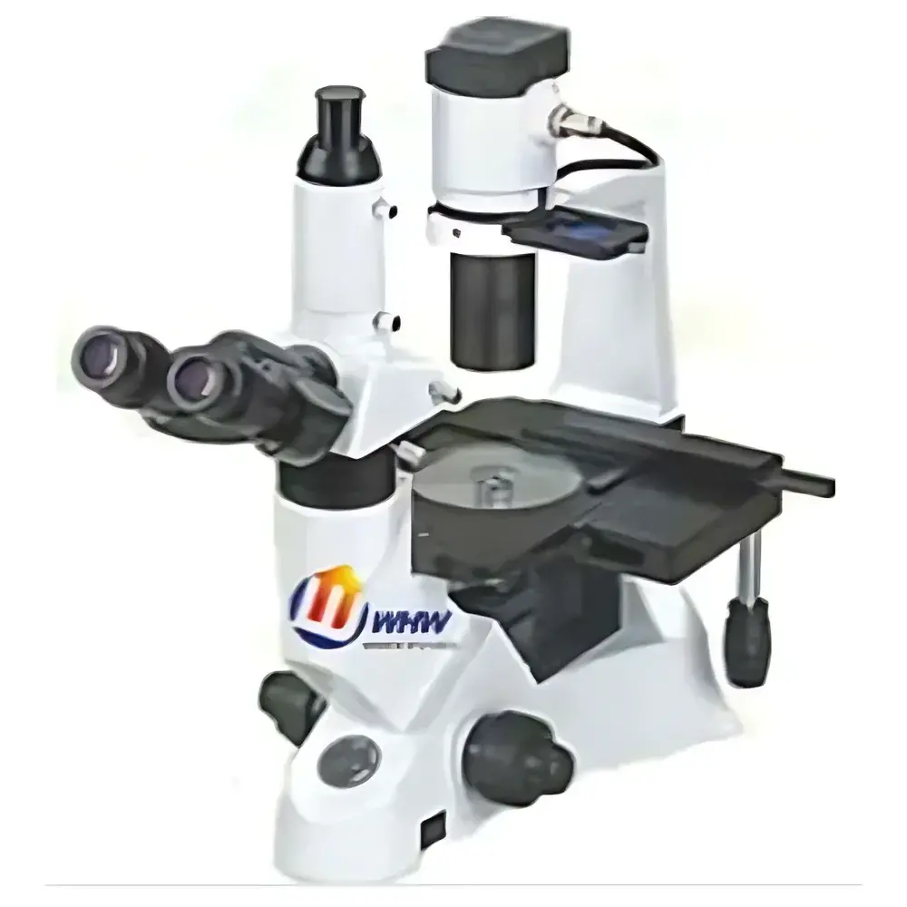
| Configuration | Trinocular |
| Observation Head | 30° inclined trinocular head, interpupillary distance adjustment range 48–75 mm |
| Eyepieces | Wide-field, high-eyepoint EW10×/22 mm |
| Objective Lenses | Infinity-corrected achromatic long-working-distance objectives — 4× (NA 0.10, WD 17.5 mm), 10× (NA 0.25, WD 8 mm), 20× (NA 0.40, WD 5.5 mm), 40× (NA 0.60, WD 3 mm) |
| Phase Contrast Objectives | PH10× (NA 0.25, WD 8 mm), PH20× (NA 0.40, WD 5.5 mm) |
| Nosepiece | Quadruple revolving nosepiece |
| Condenser | Long-working-distance condenser (NA 0.30, WD 72 mm) |
| Stage | Flat mechanical stage (160 × 250 mm) |
| Focus Mechanism | Coaxial coarse/fine focusing via nosepiece vertical movement |
| Illumination | 6 V / 30 W halogen lamp |
| Filters | 45 mm blue filter and ground glass diffuser |
| Phase Annuli | 10×–20× phase contrast annulus |
Overview
The YANRUN 37XD Inverted Biological Microscope is engineered for routine and advanced live-cell observation, tissue culture monitoring, and in vitro biological assay support. Its inverted optical configuration places the objective lenses beneath the specimen stage—enabling direct visualization of adherent or suspended cells within standard Petri dishes, multi-well plates, and flasks without specimen inversion or mounting. The system employs infinity-corrected optical design with achromatic long-working-distance (LWD) objectives, ensuring minimal thermal interference and unobstructed access to culture vessels during imaging and manipulation. Designed for robust daily use in academic teaching labs, pharmaceutical QC environments, and industrial cell biology workflows, the 37XD delivers consistent image fidelity across magnifications from 40× to 400× (with 10× eyepieces), while maintaining ergonomic operator positioning and stable mechanical alignment.
Key Features
- Trinocular observation head with 30° inclination and adjustable interpupillary distance (48–75 mm), supporting simultaneous visual observation, photomicrography, and digital imaging via C-mount adapter.
- Wide-field, high-eyepoint eyepieces (EW10×/22 mm) provide extended eye relief and full field coverage—critical for users wearing corrective eyewear and for prolonged observation sessions.
- Four standard LWD objectives (4×, 10×, 20×, 40×) with precisely calibrated numerical apertures (0.10–0.60) and working distances (3–17.5 mm) optimized for compatibility with standard thickness cultureware (e.g., 0.17 mm coverslip-corrected vessels).
- Dedicated phase contrast objectives (PH10×, PH20×) and included annular diaphragm enable label-free, high-contrast visualization of unstained, transparent living specimens—including primary neurons, fibroblasts, and epithelial monolayers—without phototoxicity.
- Removable long-working-distance condenser (NA 0.30, 72 mm WD) allows unrestricted vertical clearance up to 150 mm—accommodating tall culture vessels, perfusion chambers, or environmental control enclosures.
- Coaxial coarse/fine focusing mechanism actuates the entire nosepiece vertically, minimizing lateral drift and preserving precise focal plane registration during high-magnification work.
- Stable flat mechanical stage (160 × 250 mm) supports large-format culture dishes and enables reproducible XY positioning with dual-axis vernier scales.
Sample Compatibility & Compliance
The 37XD is validated for use with common life science consumables including 35 mm, 60 mm, and 100 mm Petri dishes; T-25, T-75, and T-175 flasks; and 6-, 12-, 24-, 48-, and 96-well plates. Its optical path accommodates standard 0.17 mm coverslip thickness and nominal vessel bottom thicknesses (0.5–1.2 mm). While not certified to ISO 13485 or FDA 21 CFR Part 11 out-of-the-box, the microscope’s mechanical stability, repeatable focus repeatability (< ±0.5 µm over 100 cycles), and traceable calibration protocol support integration into GLP-compliant workflows when paired with validated digital imaging systems and documented SOPs. It meets general safety requirements per IEC 61010-1 for laboratory electrical equipment.
Software & Data Management
The 37XD operates as a hardware platform compatible with third-party digital imaging software (e.g., NIS-Elements, ZEN Blue, CellSens, or open-source Micro-Manager) via standard C-mount interface (1× magnification, 23.2 mm thread). No proprietary firmware or closed-loop control is embedded—ensuring full user autonomy over acquisition parameters (exposure time, gain, binning, white balance) and metadata tagging. Raw image data is exported in TIFF or BMP format with embedded EXIF-like metadata (objective used, magnification, illumination intensity setting), facilitating audit-ready documentation under institutional QA/QC policies.
Applications
- Real-time monitoring of cell proliferation, migration, and confluence in monolayer cultures.
- Phase contrast assessment of embryoid body formation and stem cell differentiation kinetics.
- Quality control of hybridoma cultures and bioreactor inoculum viability.
- Support for micromanipulation procedures including microinjection, patch clamping, and laser ablation when integrated with motorized stages and environmental chambers.
- Teaching applications in undergraduate cell biology, histology, and microbiology laboratories—leveraging its mechanical simplicity, durability, and intuitive operation.
FAQ
Is the 37XD compatible with fluorescence imaging?
No—the base configuration lacks epi-illumination capability, filter cubes, or excitation light sources required for fluorescence. Fluorescence adaptation would require external modular add-ons (e.g., LED fluorescence illuminator + filter turret), which are not supported natively.
Can I upgrade to motorized focusing or automated stage control?
The 37XD features a purely manual mechanical architecture. Motorized components are not available as factory options, though third-party retrofit kits may be mechanically interfaced with appropriate engineering validation.
What is the maximum recommended specimen height with condenser removed?
Up to 150 mm—verified using standard polystyrene tissue culture flasks and glass-bottom dishes mounted on the flat stage without obstruction.
Does the microscope include camera mounting hardware?
Yes—a standard C-mount adapter (1× reduction) is supplied with the trinocular tube, enabling direct attachment of most 1/2″ or 2/3″ sensor industrial or scientific cameras.
Are replacement bulbs and fuses provided with initial shipment?
One spare 6 V / 30 W halogen bulb and two fuses are included in the accessory kit, along with alignment tools and calibration documentation.


