YANRUN FM-300 Upright Dual-Channel Epi-Fluorescence Microscope
| Brand | YANRUN |
|---|---|
| Origin | Shanghai, China |
| Manufacturer Type | Direct Manufacturer |
| Model | FM-300 Upright Dual-Channel Epi-Fluorescence Microscope |
| Optical Magnification | 40×–1000× |
| Eyepieces | Wide-Field WF10× (Φ18 mm) |
| Objective Lenses | FM-300A — PL 4×/0.10, 10×/0.25, 40×/0.65 (spring), 100×/1.25 (spring, oil) |
| Nosepiece | FM-300A — 4-position inward ball-bearing turret |
| Stage | 160 × 140 mm, mechanical movement range 75 × 50 mm |
| Condenser | Abbe N.A. 1.25 |
| Illumination | Transmitted — 6 V / 20 W halogen lamp with intensity control |
| Excitation Filter Sets | Blue — Ex 450–490 nm, Em ≥515 nm |
| Fine Focus Increment | 2 µm |
| Eyepiece Tube | Trinocular, inclined 30° |
| Focus Mechanism | Coaxial coarse/fine focusing with adjustable coarse tension, locking knob, and upper/lower limit stops |
Overview
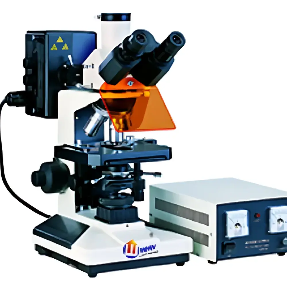
The YANRUN FM-300 Upright Dual-Channel Epi-Fluorescence Microscope is an engineered optical platform designed for routine and advanced fluorescence imaging in life science laboratories, pathology units, and materials research facilities. Built upon a rigid upright configuration, it integrates transmitted brightfield illumination with dual-band epi-fluorescence excitation—optimized for simultaneous or sequential observation of blue- and green-emitting fluorophores (e.g., DAPI/FITC, Hoechst/GFP). Its optical architecture adheres to Köhler illumination principles for both transmission and epifluorescence paths, ensuring uniform illumination, minimal stray light, and high signal-to-noise ratio. The system employs plan achromat objectives corrected for chromatic and spherical aberration across the visible spectrum, while dedicated fluorescence objectives (FL series) feature enhanced transmission in the near-UV to green region and anti-reflection coatings optimized for 365 nm, 450–490 nm, and 495–555 nm excitation bands. Mechanical stability is reinforced by a cast-metal base, precision-ground stage rails, and coaxial focusing with calibrated 2 µm fine-focus graduation—critical for Z-stack acquisition and quantitative morphometric analysis.
Key Features
- Trinocular head with 30° inclination and standardized C-mount interface for seamless integration with scientific CMOS/CCD cameras and digital imaging systems.
- Coaxial coarse/fine focusing mechanism with adjustable coarse-drive tension, mechanical upper/lower limit stops, and a 2 µm fine-focus graduation scale—enabling reproducible focal plane positioning for serial sectioning and time-lapse z-series capture.
- Dual-path illumination system: 6 V / 20 W halogen lamp with continuous intensity control for transmitted brightfield, phase contrast, and polarized light applications; separate 100 W high-pressure mercury lamp with external wide-voltage (110–220 V) power supply and optional timer function for stable, low-noise fluorescence excitation.
- Interchangeable nosepieces: FM-300A features a 4-position inward-ball-bearing turret for standard fluorescence work; FM-300B upgrades to a 5-position turret accommodating additional objectives—including phase contrast (PHP) or extended magnification options.
- Abbe condenser (N.A. 1.25) with centering screws and aperture diaphragm for precise critical illumination alignment; compatible with optional phase contrast slider assemblies (PH-I, PH-II, slide-in, or pull-out types).
- Standardized filter cube slots supporting modular excitation/emission/dichroic combinations—factory-aligned for blue (450–490 nm ex / ≥515 nm em) and green (495–555 nm ex / ≥595 nm em) channels, with optional UV (320–380 nm) and violet (380–415 nm) modules available.
Sample Compatibility & Compliance
The FM-300 accommodates standard 1″ × 3″ glass microscope slides and coverslips (0.13–0.17 mm thickness), supporting fixed and live-cell preparations, tissue sections, stained cytology smears, and semi-transparent mineral or polymer specimens. Its mechanical stage (75 × 50 mm travel) enables precise coordinate-based navigation, essential for grid-based screening and multi-field documentation. While not certified to ISO 13485 or FDA 21 CFR Part 11 out-of-the-box, the microscope’s hardware design—particularly its repeatable focus calibration, stable illumination output, and objective lens labeling per ISO 8578—supports GLP-compliant documentation workflows when paired with validated digital imaging software. All optical components comply with ISO 10934-1 (microscope nomenclature) and JIS B 7151 (objective lens marking standards). Optional phase contrast and polarizing accessories extend utility into pharmaceutical crystallography and polymer morphology studies aligned with USP and ASTM E112 methodologies.
Software & Data Management
The FM-300 interfaces via standard C-mount (0.4×, 0.5×, 1×, or 0.5× with 0.1 mm/grad reticle) and USB/VIDEO ports to third-party imaging platforms—including open-source tools (e.g., MicroManager, Fiji/ImageJ) and commercial suites (NIS-Elements, ZEN, CellSens). When used with compliant software, the system supports audit-trail-enabled image acquisition, timestamped metadata embedding (objective ID, magnification, exposure, filter set), and DICOM-SR export for integration into laboratory information management systems (LIMS). Optional 100 W constant-power mercury power supplies include built-in usage timers—facilitating lamp-hour tracking required under preventive maintenance schedules per ISO/IEC 17025 Clause 6.4.2.
Applications
- Cell biology: Immunofluorescent localization of cytoskeletal proteins (e.g., actin with phalloidin-Alexa488), nuclear counterstaining (DAPI), and co-localization studies using dual-channel overlay.
- Pathology: Rapid screening of fluorescently labeled tissue biopsies, including IHC-FISH hybrid assays on formalin-fixed paraffin-embedded (FFPE) sections.
- Microbiology: Detection of autofluorescent bacterial aggregates or GFP-expressing reporter strains in biofilm assays.
- Materials science: Visualization of fluorescent dopants in thin-film semiconductors, quantum dot dispersion homogeneity, and stress-induced birefringence in polymer laminates under combined fluorescence/polarization modes.
- Education & training: Modular configuration supports curriculum-based instruction in optical theory, fluorescence physics, and microscopy best practices per ANSI Z358.1 and CLSI H26-A4 guidelines.
FAQ
Is the FM-300 compatible with oil immersion objectives?
Yes—the 100×/1.25 NA plan achromat and fluorescence objectives are designed for use with Type A cedarwood oil (n = 1.518) and include spring-loaded mounting to protect lenses during focusing.
Can I upgrade from FM-300A to FM-300B configuration post-purchase?
The FM-300B nosepiece, stage, and condenser assemblies are mechanically and optically interchangeable with FM-300A frames; contact YANRUN technical support for retrofit kit availability and calibration verification.
What safety certifications apply to the 100 W mercury lamp power supply?
The external power supply meets IEC 61000-6-3 (EMC emission) and IEC 61000-6-2 (immunity) standards; UV-blocking housing complies with EN 62471 (photobiological safety) for Risk Group 2 classification.
Does the microscope support quantitative fluorescence intensity measurement?
Absolute quantification requires external calibration with NIST-traceable fluorescent microspheres and software-based background subtraction; the FM-300’s stable illumination and repeatable focus provide the foundational hardware consistency required for relative intensity comparisons across samples.
Are replacement filter cubes available for custom fluorophore pairs (e.g., TRITC, Cy5)?
Yes—YANRUN offers OEM-specified excitation/emission/dichroic combinations matching common fluorophores; custom cubes require minimum order quantities and 8–10 week lead time for spectral validation.


