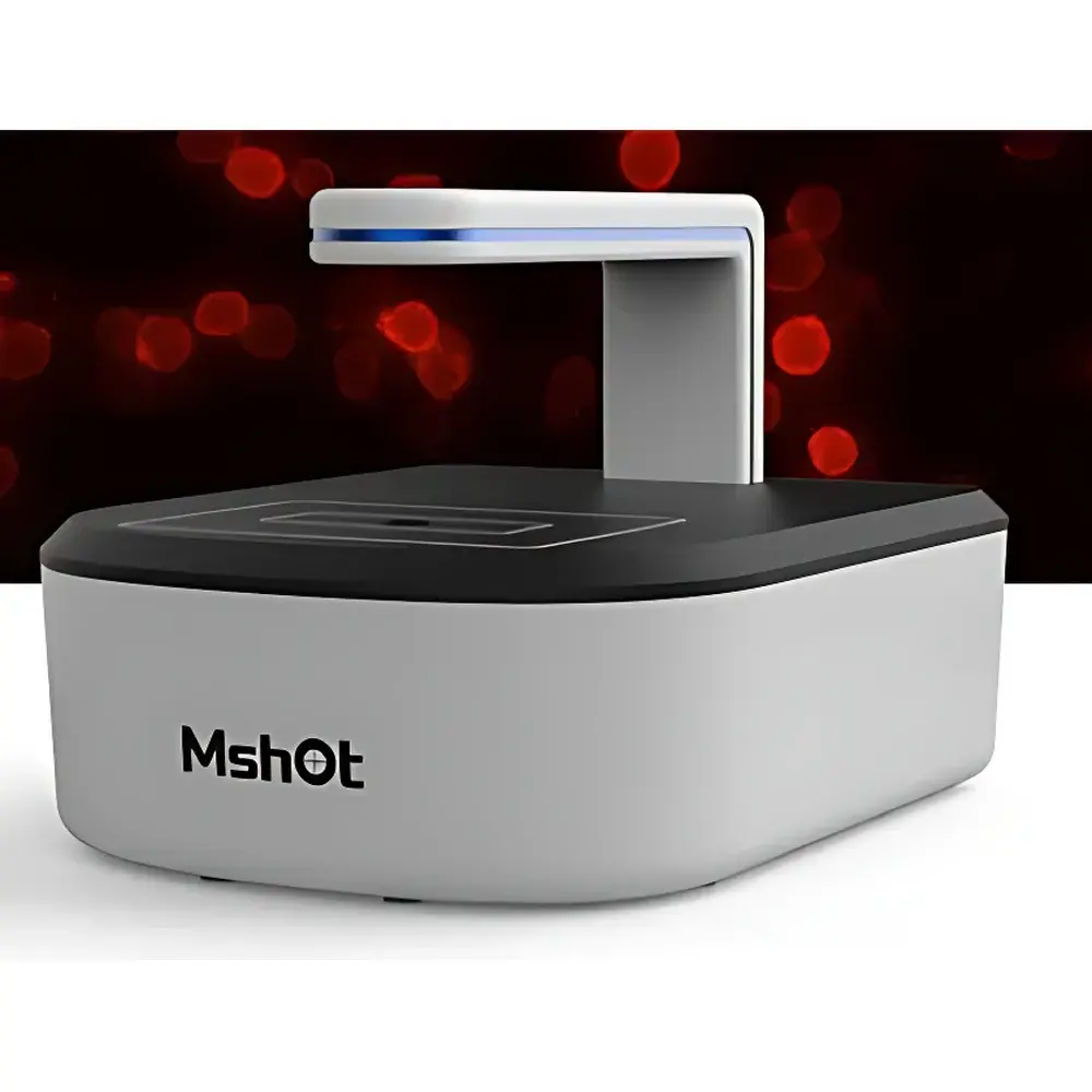
MSHOT MCS21 High-Content Live-Cell Imaging System
| Brand | MSHOT |
|---|---|
| Origin | Guangdong, China |
| Manufacturer Type | Authorized Distributor |
| Country of Origin | China |
| Model | MCS21 |
| Pricing | Upon Request |
| Imaging Modes | Brightfield, Phase Contrast, Fluorescence (Dual-Channel: Blue 475/30 nm & Green 560/40 nm) |
| Light Source | 625 nm Red LED for Phase Contrast |
| Objective | 10×/0.25 Phase Contrast |
| Z-Axis Focus | Motorized Autofocus |
| Camera | 5 MP, 2/3" CMOS, 40 fps |
| Software Architecture | Client/Server (C/S), Remote Control Enabled |
| Environmental Range | 5–42 °C, 5–95% RH |
| Power & Data | USB + DC Input |
Overview
The MSHOT MCS21 is a high-content live-cell imaging system engineered for long-term, non-invasive monitoring of adherent and suspension cultures within standard CO₂ incubators. It operates on a fixed-stage, integrated optical architecture combining transmitted brightfield, phase contrast, and dual-band fluorescence microscopy—enabling label-free morphological assessment alongside targeted fluorescent reporter tracking (e.g., GFP, RFP, Calcein-AM). Unlike conventional inverted microscopes requiring sample removal, the MCS21 mounts directly inside incubator chambers with a 70 mm working distance, minimizing thermal and pH perturbation. Its core measurement principle relies on time-lapse digital image acquisition synchronized with programmable illumination duty cycles, ensuring phototoxicity mitigation through precise LED pulse control (e.g., 625 nm red LED for phase contrast, reducing photobleaching vs. white-light sources). Designed for GLP-aligned workflows, it supports continuous acquisition over days to weeks with sub-minute temporal resolution—ideal for proliferation kinetics, confluence dynamics, wound healing assays, and transfection efficiency validation.
Key Features
- Integrated incubator-compatible design with sealed optical path and temperature-stable mechanical housing
- Dual-channel fluorescence excitation/emission filtering (475/30 nm for DAPI/GFP; 560/40 nm for RFP/mCherry) enabling multiplexed viability and localization studies
- Motorized Z-axis autofocus with closed-loop feedback, maintaining focus drift < ±0.5 µm over 72 h at 37 °C
- High-speed 5 MP CMOS sensor (2/3″ format) delivering 40 fps full-frame capture for rapid event capture (e.g., mitosis, membrane ruffling)
- Programmable LED illumination with adjustable intensity and exposure duration per channel—critical for quantitative fluorescence intensity normalization
- C/S software architecture supporting concurrent local acquisition and remote operation via secure HTTPS tunnel, including real-time preview and parameter adjustment
Sample Compatibility & Compliance
The MCS21 accommodates standard tissue-culture vessels including 6–96-well plates, chamber slides, and Petri dishes (up to 100 mm diameter). Its phase contrast optics are optimized for unstained mammalian cells (e.g., HeLa, HEK293, primary fibroblasts), while fluorescence channels support common viability dyes (propidium iodide, Hoechst), calcium indicators (Fluo-4), and genetically encoded biosensors. The system complies with IEC 61000-6-3 (EMC emission standards) and meets ISO 13485 design control principles for in vitro diagnostic support equipment. Audit trail functionality—including user login timestamps, parameter change logs, and image metadata (exposure, gain, focus position)—supports 21 CFR Part 11 readiness when deployed in regulated QC environments.
Software & Data Management
The proprietary MCS Control Suite implements a tiered analysis pipeline: raw image acquisition → background subtraction and flat-field correction → object segmentation (adaptive thresholding + watershed) → feature extraction (area, perimeter, circularity, intensity distribution). Preconfigured modules include confluence quantification (pixel-based occupancy algorithm), scratch assay migration velocity calculation (edge-tracking over time), and multi-object counting with size/gating filters. All processed data export to CSV or HDF5 formats; metadata embeds EXIF-compliant tags (DICOM-SR compatible). Remote users receive email alerts upon defined triggers (e.g., confluence >90%, focus loss >3 frames), and encrypted data sync occurs via configurable SFTP endpoints—ensuring integrity during cross-site collaboration.
Applications
- Longitudinal monitoring of CRISPR-edited cell line expansion under selection pressure
- Quantitative assessment of cytotoxicity in dose-response assays (IC₅₀ derivation from confluence decay curves)
- Real-time tracking of organelle dynamics (mitochondrial fission/fusion) using MitoTracker dyes
- Validation of siRNA knockdown efficiency via time-resolved GFP-tagged protein loss
- GMP-compliant documentation of master cell bank recovery and passage consistency
- Automated detection of mycoplasma contamination via aberrant morphology clustering in brightfield sequences
FAQ
Can the MCS21 be installed in a humidified 5% CO₂ incubator without condensation risk?
Yes—the optical head features conformal-coated PCBs and hermetically sealed lens barrels rated for continuous operation at 95% RH and 37 °C.
Does the software support batch processing of time-lapse datasets across multiple wells?
Yes—batch analysis mode applies identical segmentation and measurement parameters to all selected wells, generating consolidated Excel reports with well-position indexing.
Is calibration traceable to NIST standards?
Spatial calibration uses certified stage micrometers (NIST-traceable); intensity calibration is performed with neutral density filters and verified annually per internal SOP-IM-017.
How is data security enforced during remote access?
All remote sessions require two-factor authentication (TOTP), TLS 1.3 encryption, and role-based permissions (e.g., “Analyst” cannot modify acquisition protocols).
What maintenance intervals are recommended for sustained performance?
LED output intensity verification every 6 months; autofocus calibration check before each long-term experiment; annual preventive service includes sensor dark-frame characterization and optical alignment verification.
Related Products


