Senbe SEN785 Coherent Raman Microscopic Imaging System for Microplastic Analysis
| Brand | Senbe |
|---|---|
| Origin | Jiangsu, China |
| Manufacturer Type | Direct Manufacturer |
| Product Category | Domestic |
| Model | SEN785 |
| Instrument Type | Stimulated Raman Scattering (SRS) Microscope |
| Excitation Wavelength | 785 nm |
| Spectral Range | 200–3000 cm⁻¹ |
| Spectral Resolution | ≤8 cm⁻¹ |
| Spectral Shift Accuracy | ≤1 cm⁻¹ |
| Spectral Shift Repeatability | ≤1 cm⁻¹ |
| Detection Limit | 0.1% ethanol in N₂ |
| Dynamic Range | ≥33,000:1 |
| Detector SNR | ≥1000:1 |
| Detector | TEC-cooled back-illuminated CCD array (1024 × 58 pixels) |
| Microscope | Upright infinity-corrected plan achromat objectives (10×, 20×, 50× |
| Objective Turret | ≥4-position manual turret |
| Camera | ≥9 MP monochrome CCD |
| Stage | Mechanical XY stage (175 × 145 mm, 76 × 42 mm travel) |
| Software | Integrated spectral acquisition, multivariate analysis (PCA, PLS, CNN), HQI matching, AirPLS baseline correction, S-G/Whittaker/EMD denoising, 2D spectral mapping, and user-defined microplastic spectral library management |
Overview
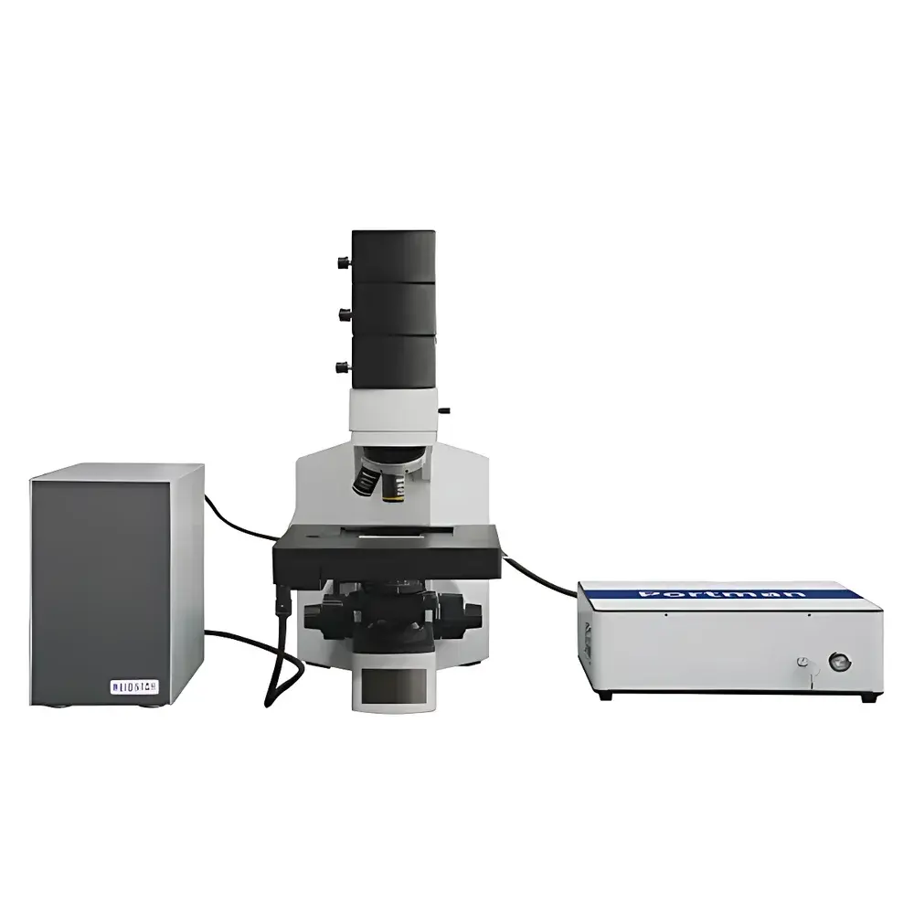
The Senbe SEN785 Coherent Raman Microscopic Imaging System is a purpose-engineered platform for label-free, chemically specific identification and spatial mapping of microplastics in complex environmental and biological matrices. Leveraging stimulated Raman scattering (SRS) as its core contrast mechanism, the system enables quantitative, non-destructive detection of polymer-based particles without staining or labeling—critical for preserving sample integrity in regulatory and research workflows. Unlike conventional fluorescence or FTIR-based methods, SRS provides high signal-to-noise vibrational contrast at video-rate acquisition speeds, making it uniquely suited for high-throughput screening of sub-100 µm particles in filter-collected water, sediment, tissue, or airborne particulate samples. The 785 nm excitation wavelength minimizes autofluorescence interference from organic matter while maintaining sufficient Raman cross-section for common polymers including polyethylene (PE), polypropylene (PP), polystyrene (PS), polyethylene terephthalate (PET), and polyvinyl chloride (PVC). The instrument integrates a high-stability upright microscope with modular laser coupling, enabling seamless transition between widefield optical imaging and confocal Raman spectral acquisition.
Key Features
- Upright infinity-corrected microscope architecture with plan achromat objectives (10×, 20×, 50× standard; optional 100× and NIR-optimized 20×/50×) for high-fidelity morphological assessment and diffraction-limited spatial registration of spectral data.
- Modular SRS optical design supporting multi-wavelength operation (532 nm, 785 nm, 1064 nm) across up to three independent detection channels—each equipped with a dedicated high-sensitivity fiber-coupled Raman probe for flexible configuration and field-deployable diagnostics.
- Pull-out mechanical coupling interface between imaging optics and Raman probe, ensuring optimal alignment stability and maximal photon collection efficiency—critical for achieving ≤8 cm⁻¹ spectral resolution and sub-0.1% concentration detection limits in aqueous suspensions.
- TEC-cooled back-illuminated CCD detector (1024 × 58 pixels) delivering ≥1000:1 signal-to-noise ratio and ≥33,000:1 dynamic range—enabling robust discrimination of weak Raman bands against heterogeneous background signals from soil, biofilm, or tissue.
- Software-controlled laser power modulation (1 mW increments), integration time tuning, and automated XY raster scanning—fully programmable for standardized method development compliant with emerging ISO/ASTM microplastic characterization guidelines.
Sample Compatibility & Compliance
The SEN785 accommodates diverse sample formats relevant to environmental monitoring and toxicological assessment: membrane-filtered water extracts (e.g., GF/F, PVDF), dried sediment smears, cryosectioned biological tissues, airborne particle filters (e.g., quartz fiber), and suspension droplets on reflective substrates. Its open optical path supports custom sample holders—including temperature-controlled stages and flow cells—for dynamic studies. From a regulatory standpoint, the system’s traceable spectral calibration (≤1 cm⁻¹ shift accuracy/repeatability), audit-ready software logging, and GLP-aligned data handling protocols align with principles outlined in ISO 21047 (microplastic identification by vibrational spectroscopy) and ASTM D8332 (standard guide for microplastic analysis in environmental media). While not FDA 21 CFR Part 11 certified out-of-the-box, the software architecture supports electronic signature implementation and full audit trail generation for GMP-aligned laboratories conducting method validation.
Software & Data Management
The integrated acquisition and analysis suite provides end-to-end workflow support—from real-time spectral preview and auto-focus-assisted targeting to multivariate classification and spatial correlation. Core capabilities include: (1) hardware-synchronized spectral acquisition with configurable averaging, dark-current subtraction, and cosmic-ray rejection; (2) advanced baseline correction using AirPLS; (3) spectral denoising via Savitzky-Golay, Whittaker, and Empirical Mode Decomposition algorithms; (4) peak detection, FWHM calculation, and integrated area quantification; (5) HQI (Hit Quality Index)-based spectral matching against reference libraries; (6) unsupervised (PCA) and supervised (PLS, CNN) machine learning models trained on user-curated microplastic spectra; and (7) 2D chemical mapping where pixel-wise Raman parameters (e.g., C–H stretch intensity, carbonyl band ratio) are rendered as pseudocolor thermograms. All raw and processed data adhere to HDF5-based storage conventions, ensuring interoperability with third-party tools such as Python-based scikit-learn or MATLAB-based Chemometrics Toolbox.
Applications
- Identification and quantification of microplastics in marine sediment cores, wastewater effluent filters, and drinking water concentrates per ISO/IEC 17025-accredited protocols.
- Spatially resolved polymer distribution mapping in histological sections of fish gills, mammalian lung tissue, or placental biopsies to assess translocation pathways.
- Discrimination of weathered vs. virgin microplastics via oxidation-sensitive Raman band ratios (e.g., C=O/C–H), supporting environmental aging studies.
- High-content screening of nanoplastic uptake in 3D organoid cultures using SRS-enabled live-cell imaging without phototoxicity.
- Development of reference spectral libraries for region-specific polymer contaminants (e.g., textile-derived acrylics, tire wear particles) under controlled inter-laboratory round-robin testing frameworks.
FAQ
What is the minimum detectable microplastic size using the SEN785 system?
The practical lower limit is governed by diffraction and signal-to-noise constraints—not instrument specification alone. With 50× objective and 785 nm excitation, lateral resolution approaches ~350 nm; however, reliable identification of particles <2 µm requires optimized filtering, background subtraction, and HQI thresholding—typically validated per ISO 21047 Annex B.
Can the system distinguish between polyethylene and polypropylene when they co-occur in mixed samples?
Yes. PE exhibits dominant peaks at ~1060 cm⁻¹ (C–C stretch) and ~1440 cm⁻¹ (CH₂ scissoring), whereas PP shows characteristic bands at ~840 cm⁻¹ (CH₃ rocking) and ~970 cm⁻¹ (isotactic helix mode). Multivariate analysis (e.g., PCA loadings or CNN feature extraction) enables robust separation even in overlapping spectral regions.
Is spectral calibration traceable to NIST standards?
The system includes built-in silicon (520.7 cm⁻¹) and cyclohexane (2847 cm⁻¹, 2910 cm⁻¹) reference standards. Users may perform additional calibration using NIST-traceable polystyrene or polyethylene film standards—procedures documented in the instrument’s calibration SOP.
Does the software support batch processing of large hyperspectral datasets?
Yes. The analysis engine supports parallelized spectral preprocessing and model inference across multi-core CPUs. Export options include CSV (peak tables), TIFF (chemical maps), and HDF5 (full cube data) for downstream statistical modeling in R or Python.
What maintenance is required for long-term spectral stability?
Annual recalibration of laser wavelength alignment and detector linearity is recommended. Optical components require periodic inspection for dust accumulation; no consumables are involved in routine operation beyond standard microscope cleaning protocols.


