SOPTOP XD Inverted Fluorescence Microscope
| Brand | SOPTOP |
|---|---|
| Origin | Zhejiang, China |
| Manufacturer Type | Direct Manufacturer |
| Regional Classification | Domestic (China) |
| Model | XD |
| Price Range | USD 14,000 – 25,500 |
| Instrument Type | Inverted Fluorescence Microscope |
| Excitation Source | High-Stability LED |
| Medical Device Certification | Not Applicable |
| Microscope Category | Conventional Fluorescence Microscope |
| Eyepieces | Wide-Field Plan Eyepieces PL10X/22 mm with Adjustable Diopter and Optional Reticle |
| Objectives | Ultra-Long Working Distance Infinity-Corrected Achromatic Objectives (4X, 10X, 20X, 40X, 60X) |
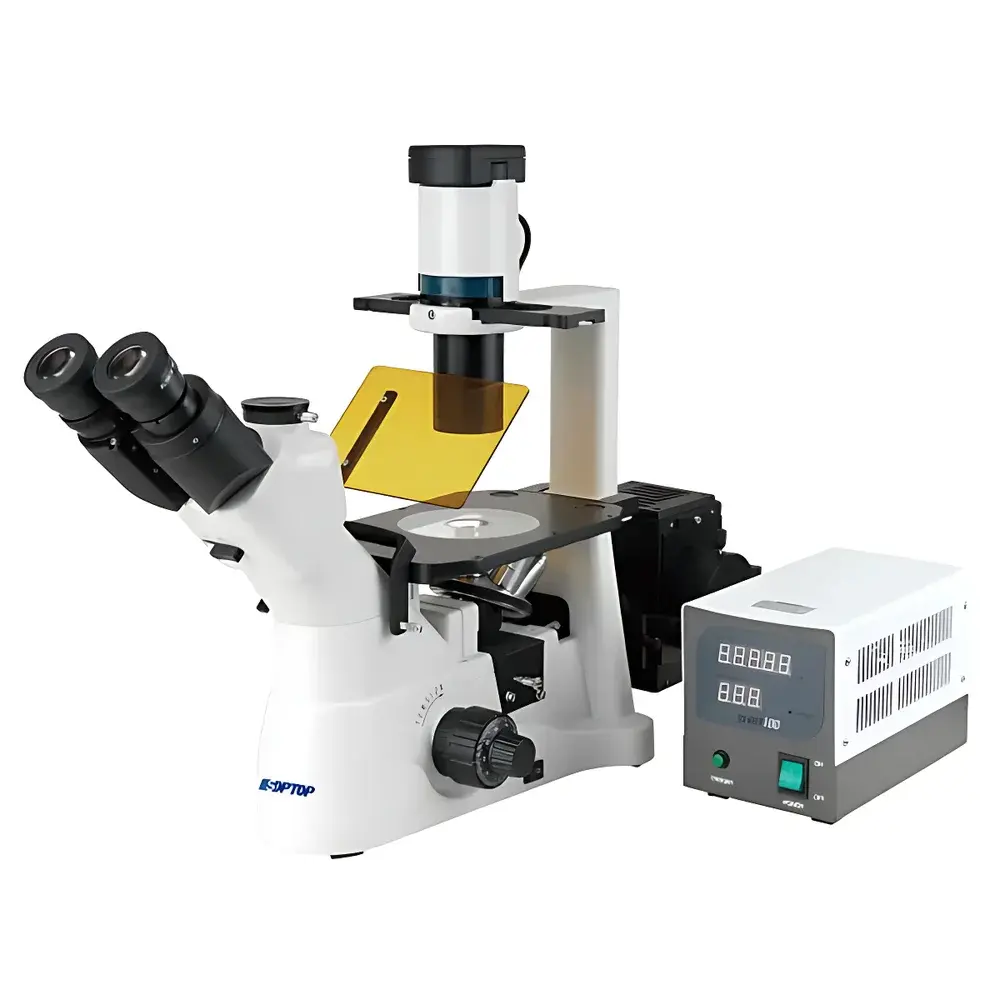
| Fluorescence Filter Cubes | B1, G1, UV1, V1 |
| Illumination | 6V/30W Halogen Lamp |
| Control Mode | Manual |
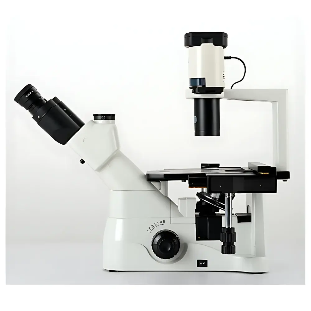
| Focus Mechanism | Coaxial Coarse/Fine Focus with Low-Position Knobs |
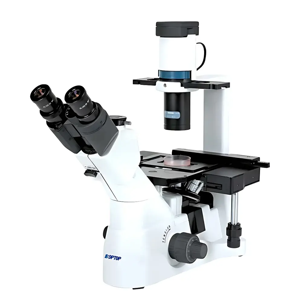
| Condenser | Removable Long Working Distance Condenser, NA 0.3, Optional Köhler or Critical Illumination |
| Observation Head | 45° Inclined Binocular or Trinocular Hinged Body |
| Stage | Fixed 250 mm × 160 mm Platform |
| Fluorescence Filters | B1/G1/UV1/V1 |
Overview
The SOPTOP XD Inverted Fluorescence Microscope is an engineered platform designed for routine and advanced live-cell observation in academic, pharmaceutical, and biotechnology laboratories. Its inverted optical architecture places the objective lenses beneath the specimen stage—enabling direct access to culture vessels such as Petri dishes, multi-well plates, and flasks without disturbing cell monolayers or suspension cultures. The system operates on standard widefield fluorescence principles: excitation light (delivered via high-stability LEDs and/or a 6V/30W halogen lamp) passes through dichroic mirrors and bandpass filters to selectively illuminate fluorophores; emitted fluorescence is then separated from excitation light by emission filters and directed to the eyepiece or camera port. This configuration supports both transmitted-light techniques—including brightfield, phase contrast, and differential interference contrast—and epifluorescence imaging across UV–visible spectral bands (B, G, UV, and V filter sets). The XD’s optical path is fully infinity-corrected, ensuring compatibility with modern digital imaging systems and minimizing chromatic aberration across magnifications.
Key Features
- Ultra-long working distance (ULWD) infinity-corrected objectives—up to 12 mm at 40X—accommodate thick culture vessels and environmental chambers without physical interference.
- Dual illumination capability: selectable Köhler or critical illumination modes via a rotatable condenser mount; includes field and aperture diaphragms for precise contrast and resolution control.
- Modular fluorescence system featuring four standardized filter cubes (B1, G1, UV1, V1) with high-transmission multilayer dielectric coatings, steep cut-on/cut-off edges (>99% blocking outside passband), and minimal autofluorescence background.
- Semi-apochromatic plan fluorescence phase contrast objectives (20X, 40X) deliver enhanced color correction and numerical aperture (NA ≥ 0.75), improving signal-to-noise ratio and axial resolution in dual-mode (fluorescence + phase) imaging.
- Ergonomic manual operation: coaxial coarse/fine focus with torque-adjustable fine-tuning (1 µm graduation), low-hand-position knobs, and 45° inclined trinocular head compatible with C-mount adapters for sCMOS or EMCCD cameras.
- Expandable mechanical stage system supporting ISO-standard tissue culture formats: 6-/12-/24-/96-well plates, Φ35 mm Petri dishes, Teraseki slides, and custom inserts via slotted metal or glass stage plates.
Sample Compatibility & Compliance
The XD microscope accommodates a broad range of biological specimens—from adherent mammalian cell lines (e.g., HeLa, NIH/3T3) and primary neurons to zebrafish embryos, organoids, and co-cultures in hydrogel matrices. Its long working distance optics and open-stage design facilitate integration with environmental control units (temperature, CO₂, humidity) for time-lapse experiments up to 72 hours. While not certified as a medical device under FDA 21 CFR Part 820 or EU MDR, the instrument conforms to IEC 61000-6-3 (EMC emissions) and IEC 61010-1 (safety for laboratory equipment). All optical components meet ISO 10110 surface quality standards (scratch-dig 60–40), and fluorescence filter spectral data are traceable to NIST-calibrated spectrophotometers. Documentation supports GLP-compliant recordkeeping when paired with validated image acquisition software.
Software & Data Management
The XD interfaces with third-party imaging platforms including NIS-Elements (Nikon), ZEN Blue (Zeiss), and open-source tools such as Micro-Manager and Fiji/ImageJ via USB 3.0 or GigE Vision-compatible cameras. Optional SOPTOP-provided acquisition software offers synchronized multi-channel capture, Z-stack reconstruction, time-lapse scheduling, and basic intensity profiling. Audit trail functionality—including user login timestamps, parameter change logs, and metadata embedding (EXIF/OME-TIFF)—supports alignment with FDA 21 CFR Part 11 requirements when deployed in regulated QC environments. Raw image files retain full bit-depth (12–16 bit) and spatial calibration metadata for downstream quantitative analysis (e.g., colocalization, FRET, morphometric segmentation).
Applications
- Live-cell monitoring of proliferation, migration, and apoptosis using fluorescent dyes (e.g., Calcein-AM/PI, Hoechst 33342, CellTracker dyes).
- Transfection efficiency assessment via GFP/RFP-tagged constructs and subcellular localization studies (e.g., mitochondrial, nuclear, lysosomal targeting).
- Phase contrast–guided micromanipulation prior to fluorescence imaging—ideal for single-cell picking or microinjection workflows.
- Quality control of stem cell differentiation batches using morphological criteria and lineage-specific reporter expression.
- Preclinical assay development for drug screening, including cytotoxicity assays (e.g., LDH release) and receptor internalization kinetics.
- Teaching laboratories requiring robust, serviceable instrumentation for undergraduate microscopy training in cell biology and histology.
FAQ
Is the XD microscope compatible with motorized Z-axis or autofocus systems?
No—the XD is a manually operated platform. Motorized focus, stage, or filter cube turrets are not supported natively but may be integrated externally via third-party OEM modules (e.g., Prior Scientific ProScan III) using TTL or RS-232 triggers.
Can the halogen lamp be replaced with an LED-based transmitted-light source?
Yes—SOPTOP offers optional LED illuminators (e.g., SOPTOP L-LED6000) with adjustable intensity and CCT tuning (5000–6500 K), enabling stable white-light illumination without thermal drift or bulb replacement cycles.
What is the maximum usable magnification with the 60X ULWD objective?
At 60X with PL10X eyepieces, total magnification is 600X; with optional PL15X eyepieces, it reaches 900X. However, practical resolution remains limited by NA (0.75) and visible-light wavelength (~260 nm lateral resolution per Rayleigh criterion).
Are fluorescence filter cubes available for DAPI/FITC/TRITC/Cy5 configurations?
Standard B1/G1/UV1/V1 cubes cover common fluorophores, but custom cubes for DAPI/FITC/TRITC/Cy5 are available upon request with lead times of 6–8 weeks and spectral validation reports.
Does SOPTOP provide application support or training for new users?
Yes—SOPTOP offers remote video-based setup assistance, protocol optimization guidance, and on-site training (fee-based) covering optical alignment, Köhler illumination calibration, and fluorescence quantification best practices.


