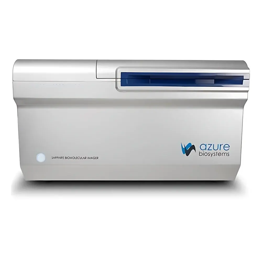
Azure Biosystems Sapphire RGBNIR Laser Scanning Imaging System
| Brand | Azure Biosystems |
|---|---|
| Origin | USA |
| Model | Sapphire RGBNIR |
| Instrument Type | Multicolor Fluorescent Gel Imaging System |
| CCD Resolution | 6.1 MP |
| Bit Depth | 16-bit |
| Dynamic Range | ≥6 OD |
| CCD Sensor Size | Large-format CCD |
| Detection Sensitivity | fg-level |
| Signal-to-Noise Ratio | High SNR |
| Optical Lens | Motorized Autofocus |
| Excitation Sources | Four Solid-State Lasers (RGB + NIR) |
| Detector Architecture | Integrated PMT, APD, and CCD Detection Modalities |
| Imaging Modes | RGB Fluorescence, NIR Fluorescence, Chemiluminescence, Visible Light, Phosphor Imaging (with optional PI module), Autoradiography |
Overview
The Azure Biosystems Sapphire RGBNIR Laser Scanning Imaging System is a high-performance, multi-modal detection platform engineered for quantitative molecular imaging across life science laboratories. Unlike conventional CCD-based gel documentation systems, the Sapphire RGBNIR employs a precision laser scanning architecture—utilizing four independently addressable solid-state lasers (473 nm, 532 nm, 635 nm, and 785 nm)—to deliver spatially resolved excitation with minimal photobleaching and optimal signal specificity. Its hybrid detection system integrates three complementary sensor technologies—photomultiplier tubes (PMTs) for ultra-low-light chemiluminescence and phosphor imaging, avalanche photodiodes (APDs) for high-speed NIR fluorescence quantification, and a large-format, cooled 6.1-megapixel CCD for high-resolution visible and RGB fluorescence capture. This tri-detection architecture enables true simultaneous or sequential multi-channel acquisition without optical crosstalk, supporting rigorous quantitative comparisons across diverse sample types and labeling chemistries.
Key Features
- Laser-scanning optics with motorized autofocus and precision stage positioning ensure consistent pixel registration and reproducible image geometry across repeated acquisitions.
- Dynamic range exceeding 6 optical density (OD) units—validated per ISO 15739—enables accurate quantification of both weak and saturated signals within a single scan, eliminating the need for exposure bracketing.
- 10 µm spatial resolution at native scan mode, achieved through diffraction-limited laser spot size and sub-pixel scanning interpolation, supports detailed analysis of fine electrophoretic bands and microarray features.
- fg-level chemiluminescent detection sensitivity—demonstrated using horseradish peroxidase (HRP)-conjugated antibodies on nitrocellulose membranes—meets or exceeds performance benchmarks defined in ASTM E2912 for low-abundance protein detection.
- Modular detector configuration allows field-upgradable integration of the optional Phosphor Imaging (PI) module, enabling compliant autoradiographic analysis of 32P, 33P, 125I, and 35S isotopes under ALARA principles.
- Fully automated calibration routines—including flat-field correction, laser power stabilization, and detector gain normalization—are executed prior to each imaging session to maintain inter-run traceability.
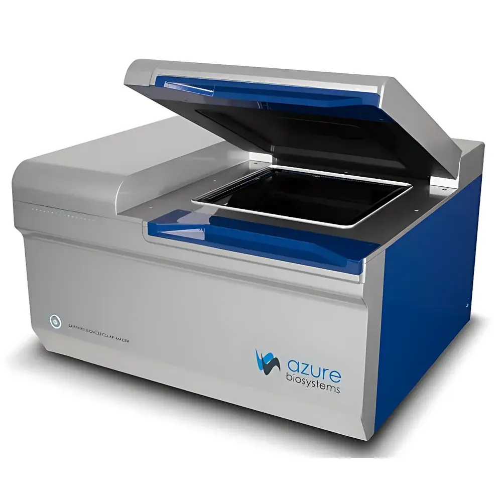
Sample Compatibility & Compliance
The Sapphire RGBNIR accommodates standard electrophoresis formats including mini- and midi-gels (up to 20 × 20 cm), blot membranes (PVDF, nitrocellulose, nylon), glass slides (for tissue sections and arrays), and microplates (96- and 384-well). All imaging modes comply with GLP and GMP documentation requirements: audit trails record operator ID, timestamp, instrument configuration, exposure parameters, and post-acquisition processing steps. Software-generated reports include metadata compliant with MIAME and MIAPE standards for proteomics and genomics data submission. The system supports 21 CFR Part 11–compliant electronic signatures when deployed with AzureSpot™ Enterprise software and validated network infrastructure.
Software & Data Management
Controlled by AzureSpot™ Imaging Software (v7.x), the platform provides a unified interface for acquisition, visualization, and analysis. The software implements non-linear intensity scaling algorithms optimized for wide-dynamic-range data, along with background subtraction models based on rolling-ball and morphological filtering. Quantitative modules include band/spot volume integration, ratio-metric multi-channel normalization, and statistical comparison across replicates using ANOVA or t-test frameworks. Raw image data are stored in vendor-neutral TIFF format with embedded EXIF and custom XML metadata; processed results export to CSV, PDF, or publication-ready PNG/SVG. Local database archiving supports version-controlled experiment tracking, while optional AzureCloud™ integration enables secure remote access and collaborative annotation.
Applications
The Sapphire RGBNIR serves as a central imaging hub for quantitative workflows across core facilities and research labs. It is routinely deployed for fluorescent Western blotting (including In-Cell and In-Gel variants), multiplexed NIR immunoassays (e.g., LI-COR-compatible assays), two-dimensional electrophoresis (2-DE) spot matching, DNA fragment analysis (ethidium bromide, SYBR Safe, GelRed), Coomassie- and silver-stained gels, nucleic acid microarrays, protein microarrays, and formalin-fixed paraffin-embedded (FFPE) tissue section imaging. Its ability to resolve spectrally distinct fluorophores—such as Alexa Fluor 488/647/790 and IRDye 680/800—without filter wheel switching ensures experimental throughput and minimizes user-induced variability.
FAQ
What regulatory standards does the Sapphire RGBNIR support for regulated environments?
The system meets ISO/IEC 17025 requirements for testing laboratories and supports full 21 CFR Part 11 compliance when configured with AzureSpot™ Enterprise, including electronic signatures, audit trails, and role-based access control.
Can the Sapphire RGBNIR perform quantitative comparisons between chemiluminescent and fluorescent signals from the same blot?
Yes—sequential scanning under identical geometric conditions, coupled with calibrated detector response curves, enables cross-modal normalization using internal reference standards such as total protein staining or housekeeping gene probes.
Is the 6.1 MP CCD used for all imaging modalities, or only specific ones?
The CCD is dedicated to high-resolution visible-light, RGB fluorescence, and stained-gel imaging; PMT and APD detectors handle low-light chemiluminescence and NIR fluorescence, respectively, optimizing signal-to-noise for each modality.
How is laser alignment and optical stability maintained over time?
The system incorporates an internal reference photodiode array and closed-loop feedback during warm-up and idle cycles, with automatic recalibration triggered after every 24 hours of operation or following environmental temperature shifts >2°C.
Does the platform support third-party analysis software via file export?
All raw images export as 16-bit TIFF files with complete metadata headers; compatible with ImageJ/Fiji, MATLAB, Python (via tifffile and ome-tiff libraries), and commercial packages including GeneTools and Quantity One.


