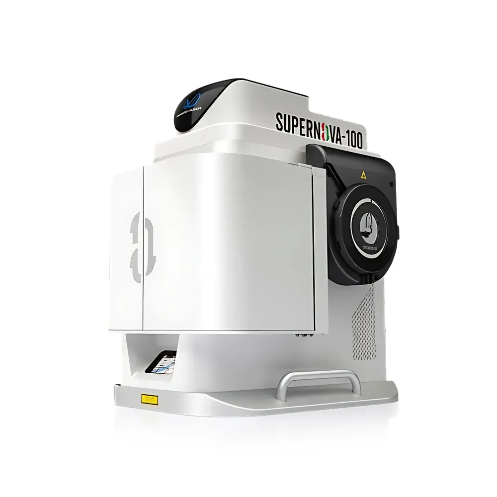
Transcend Vivoscope SUPERNOVA-100 Miniaturized Two-Photon Microscope
| Brand | Transcend Vivoscope |
|---|---|
| Model | SUPERNOVA-100 |
| Probe Mass | 2.6 g |
| Lateral Resolution | 0.65 µm |
| Field of View | 1.0 mm × 0.87 mm |
| Imaging Depth | up to 800 µm |
| Laser Compatibility | Standard Femtosecond Laser Interfaces (e.g., Coherent Chameleon, Spectra-Physics Mai Tai) |
| Multi-Modal Integration | EEG, EMG, DBS, Calcium Indicators (e.g., GCaMP), ACh Sensors (e.g., GACh), Flexible Hydrogel Electrodes (MTEHy) |
Overview
The Transcend Vivoscope SUPERNOVA-100 is an integrated, miniaturized two-photon microscope engineered for high-fidelity in vivo neural imaging in freely behaving mammals. Unlike conventional benchtop two-photon systems, the SUPERNOVA-100 implements a fully integrated optical-mechanical-electronic architecture centered on the FHIRM-series microprobe—a 2.6 g head-mounted imaging module that maintains diffraction-limited resolution while enabling unrestricted locomotion. Its core optical design leverages near-infrared (NIR) femtosecond excitation (typically 920–1300 nm) to achieve nonlinear two-photon fluorescence excitation, minimizing scattering and phototoxicity in deep cortical and subcortical tissues. The system supports simultaneous volumetric acquisition across millimeter-scale fields of view with submicron lateral resolution (0.65 µm), permitting stable, long-term tracking of dendritic spines, axonal boutons, and somatic calcium dynamics at depths exceeding 800 µm in murine cortex and hippocampus. Designed for rigorous neuroscience workflows, it operates under standard laboratory environmental conditions without requiring vibration isolation tables or climate-controlled enclosures.
Key Features
- Ultra-lightweight, head-mounted probe: 2.6 g mass ensures minimal behavioral perturbation in mice and rats; ergonomic mounting interface compatible with standard stereotaxic adapters and custom skull fixtures.
- High-resolution, large-field imaging: Achieves 0.65 µm lateral resolution with 1.0 mm × 0.87 mm field of view—enabling concurrent monitoring of >1,000 neurons within a single frame.
- Deep-tissue penetration: Optimized scan optics and dispersion-compensated beam delivery support robust signal acquisition from Layer VI cortex to hippocampal CA1 and striatal regions (up to 800 µm depth).
- Modular laser integration: Native compatibility with commercial femtosecond oscillators (e.g., Coherent Chameleon Vision-S, Spectra-Physics InSight X3) via standardized fiber-coupled input and real-time dispersion management.
- Multi-modal synchronization: TTL-triggered hardware interfaces for precise temporal alignment with electrophysiology (EEG/EMG), optogenetic stimulation, deep brain stimulation (DBS), and behavioral tracking systems.
- Robust mechanical architecture: Monolithic aluminum chassis with thermal-stable optical path; no moving parts in the probe head; designed for repeated sterilization and multi-week chronic implantation studies.
Sample Compatibility & Compliance
The SUPERNOVA-100 is validated for longitudinal in vivo imaging in transgenic and virally transduced rodents (C57BL/6, Thy1-GCaMP6s, Ai95, vGlut1-Cre, PV-Cre, VIP-Cre lines). It accommodates both cranial window preparations (glass or polymer-based) and thinned-skull protocols. All optical components comply with IEC 60825-1:2014 Class 1 laser safety standards when operated within specified power limits. The system supports GLP-compliant data acquisition through timestamp-synchronized metadata logging (frame time, laser power, scan parameters, trigger events). While not FDA-cleared for human use, its design aligns with ISO 13485 principles for research-grade instrumentation and meets essential requirements for preclinical neuroimaging under AAALAC-accredited animal facilities.
Software & Data Management
Acquisition is controlled via the proprietary VivoScan Suite, a MATLAB-based platform offering real-time preview, adaptive scanning (ROI selection, z-stack automation, resonant vs. galvo modes), and on-the-fly motion correction using cross-correlation algorithms. Raw data are saved in standardized HDF5 format with embedded metadata (including NIH ImageJ-compatible headers), ensuring interoperability with open-source analysis pipelines (CaImAn, Suite2p, SIMA). The software supports audit-trail logging per FDA 21 CFR Part 11 guidelines—including user authentication, parameter change history, and electronic signature capture—making it suitable for regulated pharmacology and translational neuroscience studies. Export modules enable direct integration with Python, R, and commercial platforms such as NeuroExplorer and Spike2.
Applications
- Mapping dynamic microcircuit motifs during social competition (e.g., dmPFC VIP→PV→PYR disinhibitory motifs, as reported in Neuron, 2022)
- Longitudinal calcium imaging of spinal cord circuits during itch-evoked scratching behavior (National Science Review, 2022)
- Single-trial acetylcholine release detection across multiple brain regions using genetically encoded ACh sensors (Nature Methods, 2020)
- Chronic imaging of dorsal horn neurons during neuropathic pain progression (bioRxiv, 2022)
- Correlating GABAergic Ca2+ transients in prelimbic cortex with social interaction metrics (Science Advances, 2022)
- Spatiotemporal characterization of seizure onset and propagation via simultaneous mTPM + EEG (Neuroscience Bulletin, 2022)
- Functional validation of flexible hydrogel-based neural interfaces (e.g., MTEHy electrodes) in freely moving subjects (Acta Biomaterialia, 2022)
FAQ
Is the SUPERNOVA-100 compatible with non-murine models?
Yes—the probe’s adjustable mounting geometry and lightweight design support adaptation to rats, ferrets, and juvenile non-human primates, subject to institutional IACUC approval and custom fixture development.
What calcium indicators are validated for use with this system?
GCaMP6f/s, jGCaMP8, RCaMP, XCaMP, and synthetic dyes including Cal-520 AM and OGB-1 AM have been successfully imaged under standard excitation wavelengths (920 nm, 1040 nm, and 1300 nm).
Does the system support volumetric (3D) imaging?
Yes—via piezo-driven objective lens translation or remote focusing modules (optional add-on), enabling rapid z-stack acquisition at frame rates up to 30 Hz for 16-plane volumes.
Can the probe be sterilized between subjects?
The titanium-alloy probe housing and sapphire window are autoclavable (121°C, 20 min); optical fibers require ethanol wipe-down and UV-C exposure per manufacturer protocol.
Is technical support available for custom experimental integration?
Transcend Vivoscope provides application engineering support—including TTL interface schematics, LabVIEW drivers, and Python API documentation—for integrating third-party hardware and developing bespoke behavioral paradigms.


