Artinis CDMAM 4.0 Breast X-ray Contrast-Detail Phantom and Analysis Software
| Brand | Artinis |
|---|---|
| Origin | Netherlands |
| Model | CDMAM 4.0 |
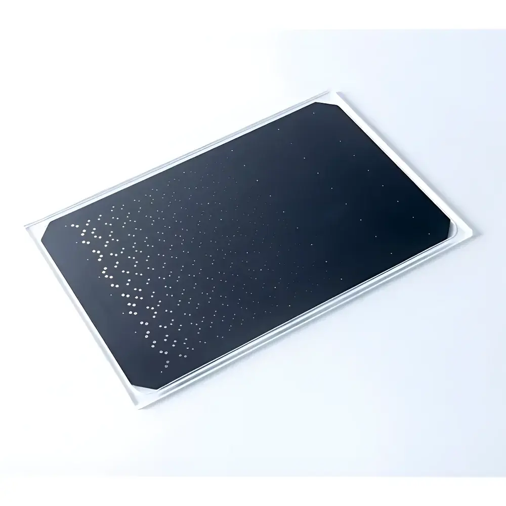
| Phantom Dimensions | 240 × 162 × 3 mm (PMMA cover), 0.5 mm polished aluminum (99.5%) base |
| Gold Disc Array | 672 discs across 21 size steps (diameters: 0.08–2.0 mm), 16 thickness levels per diameter |
| Reference Discs | 43 additional calibration discs |
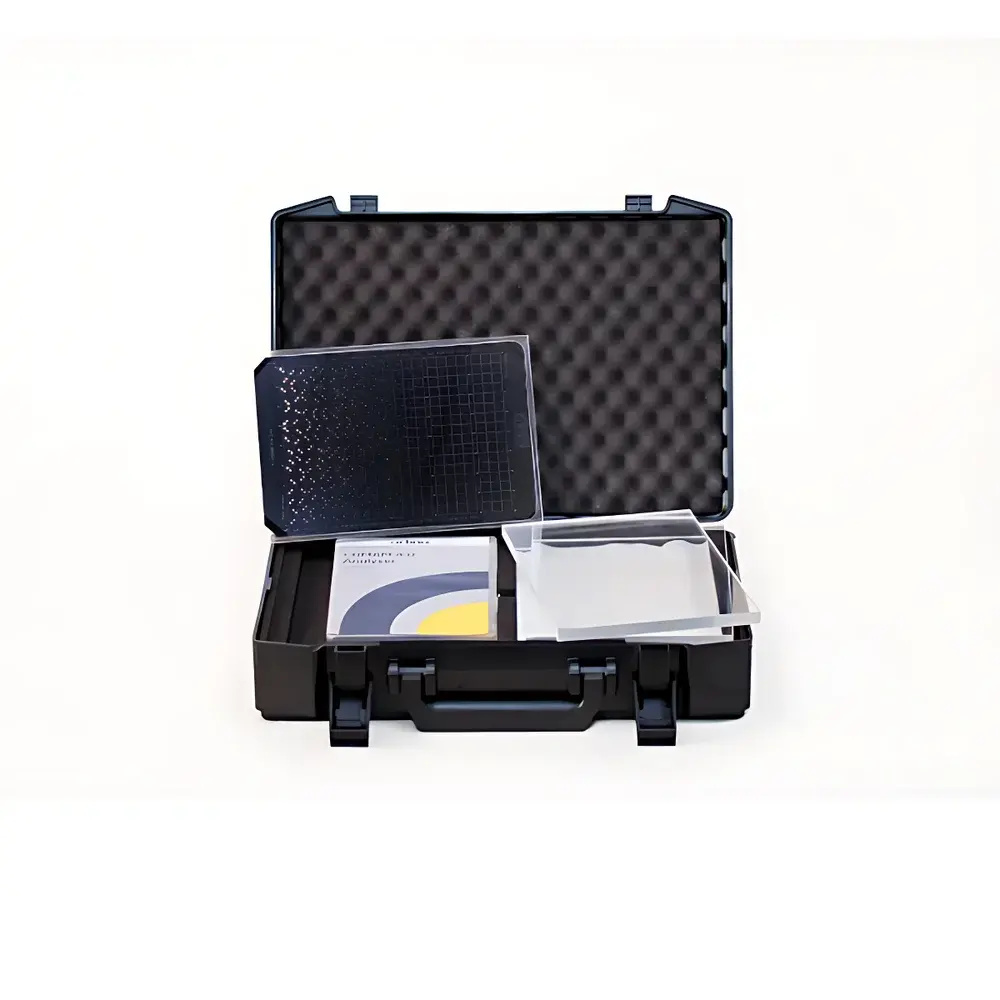
| PMMA Plates Included | 4 × 10 mm (surface polish, thickness tolerance ±0.1 mm) |
| Software | CDCOM-based CDMAM 4.0 Image Analysis Software (v4.x), DICOM-compatible, Windows 7/8/10, USB port required |
| Compliance | EU Guidelines for Quality Assurance in Breast Cancer Screening and Diagnosis (4th ed.), NHSBSP Equipment Report 0604 (3rd ed., Apr 2009), IEC 61223-3-2, EUREF CDCOM protocol |
Overview
The Artinis CDMAM 4.0 Breast X-ray Contrast-Detail Phantom and Analysis Software is a metrologically traceable reference standard engineered for quantitative performance evaluation of digital mammography systems. It implements the psychophysical principle of threshold detection—measuring the minimum contrast required to reliably identify gold disc targets of defined diameters and thicknesses under controlled exposure conditions. Designed in strict alignment with the European Guidelines for Quality Assurance in Breast Cancer Screening and Diagnosis (4th edition), the phantom enables objective assessment of system spatial resolution, low-contrast detectability, and dose-response behavior. Its construction follows ISO 12233-derived tolerancing principles for geometric fidelity and material homogeneity, ensuring reproducible test conditions across clinical sites and accreditation audits. Unlike legacy phantoms, CDMAM 4.0 integrates physical design and algorithmic analysis into a single validated workflow—eliminating inter-observer variability while supporting automated, audit-ready reporting.
Key Features
- Precision-engineered 0.5 mm thick polished aluminum (99.5% purity) base providing uniform X-ray attenuation and minimal scatter contribution.
- 672 gold discs distributed across 21 logarithmic diameter steps (0.08 mm to 2.0 mm), each with 16 optimized gold thickness levels—enabling granular mapping of the contrast-detail function (CDF).
- 43 supplementary reference discs positioned outside the primary test field to calibrate and validate automated scoring algorithms.
- Included set of four 10 mm PMMA plates (±0.1 mm thickness tolerance, optically polished surfaces) for beam hardening simulation and kVp-dependent contrast evaluation.
- Fully DICOM-compliant analysis software built upon the EUREF CDCOM framework—certified for use in GLP/GMP-aligned QA programs and FDA 21 CFR Part 11–compliant environments when deployed with appropriate audit-trail configuration.
- Automated psychometric curve generation using probit analysis; calculates threshold contrast-thickness values per disc size in accordance with Annex B of the EU QA Guidelines 4th edition.
Sample Compatibility & Compliance
The CDMAM 4.0 phantom is compatible with all full-field digital mammography (FFDM) systems, including those equipped with slot-scan, CCD, or flat-panel detectors. It supports both conventional 2D acquisition and digital breast tomosynthesis (DBT) mode testing when used with appropriate acquisition protocols (e.g., single projection DBT slices). The phantom conforms to IEC 61223-3-2 for acceptance and constancy testing of medical X-ray equipment. Its design satisfies the technical requirements outlined in the NHSBSP Equipment Report 0604 (3rd edition) for commissioning and routine quality control. All materials—including the 99.5% aluminum base and medical-grade PMMA cover—are certified for radiographic stability and long-term dimensional integrity under repeated irradiation.
Software & Data Management
The CDMAM 4.0 Analysis Software implements the EUREF CDCOM methodology with enhancements for batch processing, longitudinal trend analysis, and multi-system comparison. Input requires only standard DICOM image files (no proprietary format conversion). Each analysis generates a comprehensive report containing: IQF−1 (Inverse Quality Factor), total detected gold disc count (%), psychometric curves (probit-transformed detection probability vs. contrast-thickness), individual CD curves per diameter step, and deviation maps relative to historical baselines. Reports are exportable as PDF and CSV for integration into enterprise QC databases. The software maintains full audit trails—including user login timestamps, parameter modifications, and version-controlled analysis logs—supporting compliance with ISO 13485 and FDA 21 CFR Part 11 when operated on validated Windows platforms with appropriate access controls.
Applications
- Quantitative constancy testing of detector MTF, noise power spectrum (NPS), and detective quantum efficiency (DQE) via contrast-detail modeling.
- Determination of optimal kVp/mAs combinations for specific breast thicknesses (simulated via PMMA plate stacking).
- Inter-system comparison during procurement, site commissioning, or multi-center trials—enabling normalized performance benchmarking.
- Validation of AI-based CADe/CADx algorithms by providing ground-truth detectability thresholds for training data annotation.
- Supporting regulatory submissions (e.g., CE marking, FDA 510(k)) through documented evidence of imaging system sensitivity and reproducibility.
- Training radiographers and medical physicists in standardized QA methodology aligned with European Reference Centers (EUREF) best practices.
FAQ
Is the CDMAM 4.0 phantom compatible with digital breast tomosynthesis (DBT) systems?
Yes—when acquiring individual DBT projection images or reconstructed slices at appropriate magnification and detector binning, the phantom provides valid contrast-detail metrics for system-level optimization.
Does the analysis software require a dedicated license server or network installation?
No—CDMAM 4.0 Analysis Software operates as a standalone Windows application; licensing is node-locked to the host machine via USB dongle or hardware key.
Can the software process images from non-Artinis mammography systems?
Yes—the software accepts any DICOM-compliant mammographic image containing the full phantom field-of-view and appropriate header metadata (e.g., pixel spacing, kVp, mAs, target/filter).
How frequently should the phantom be recalibrated or replaced?
The phantom requires no periodic recalibration; its physical specifications are inherently stable. However, visual inspection for disc adhesion integrity and surface scratches is recommended before each use per ISO 15708-2 guidance.
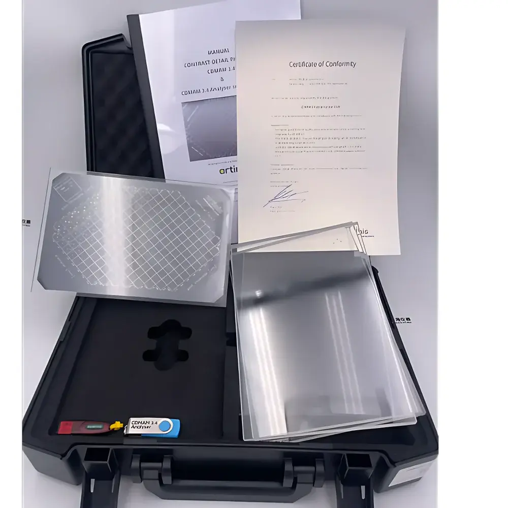
Does the package include documentation compliant with ISO/IEC 17025 for accredited laboratories?
Yes—each unit ships with a manufacturer’s certificate of conformance, traceable to NPL (UK) and VSL (NL) reference standards, along with a detailed uncertainty budget for disc diameter and thickness tolerances.