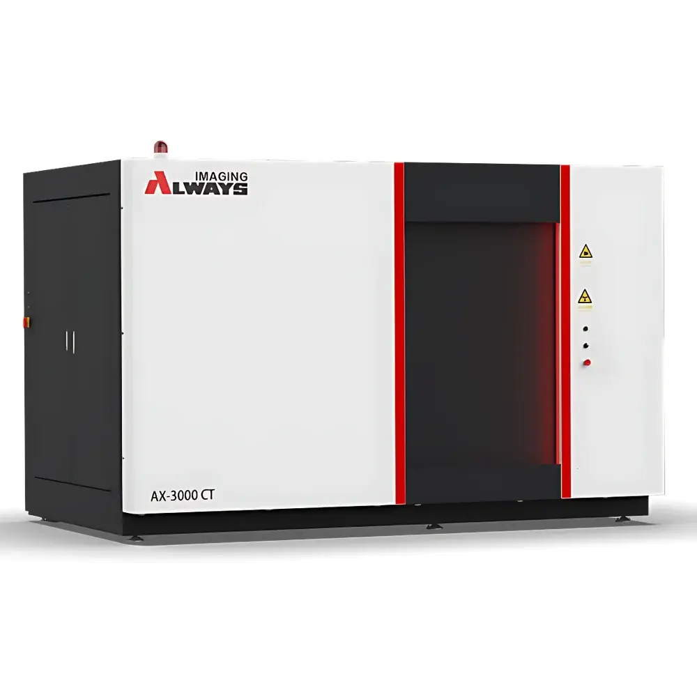
Always Imaging AX-3000CT-D Dual-Source Micro-Nano Industrial Computed Tomography System
| Brand | Always Imaging |
|---|---|
| Origin | Zhejiang, China |
| Manufacturer Type | Direct Manufacturer |
| Origin Category | Domestic |
| Model | AX-3000CT-D |
| Detector Type | Digital Flat-Panel Detector |
| Scanning Mode | Translation-Rotation (TR) |
| Spatial Resolution | 1.5–2 µm / 3–4 µm |
| Density Resolution | <1% |
| X-ray Energy | 160–300 kV (dual-source configurable: e.g., 180 kV + 240 kV) |
| Maximum Sample Dimensions | Ø600 mm × H1000 mm |
| Sample Weight Capacity | 50 kg / 100 kg / 200 kg |
| System Footprint | 3500 mm × 2000 mm × 2300 mm |
| Focal Spot Size | ≤1.5 µm (nano-focus mode, 180 kV) / ≤4 µm (micro-focus mode, 240–300 kV) |
| Detector Pixel Pitch | 100–200 µm |
| Detector Array Size | Up to 3072 × 3072 pixels |
| Field of View | Up to 427 mm × 427 mm |
| Radiation Shielding | Integrated walk-in lead-lined room (<1 µSv/h ambient dose rate) |
Overview
The Always Imaging AX-3000CT-D is a dual-source micro-nano industrial computed tomography (CT) system engineered for high-fidelity, non-destructive 3D volumetric imaging of complex engineering components and scientific specimens. It operates on the principle of cone-beam X-ray computed tomography, where two independently controllable X-ray sources—each optimized for distinct energy ranges and focal spot sizes—are paired with a high-dynamic-range digital flat-panel detector. This architecture enables adaptive source selection or simultaneous dual-energy acquisition, facilitating material discrimination, artifact suppression, and enhanced contrast-to-noise ratio in heterogeneous samples. Designed as a large-format, walk-in CT platform, the AX-3000CT-D integrates a fully shielded lead-lined enclosure compliant with GBZ 130–2020 (Chinese National Standard for Medical and Industrial X-ray Equipment Protection) and IEC 61331-1:2014. Its TR (translation-rotation) scanning geometry ensures mechanical stability and geometric fidelity over extended scan trajectories—critical for maintaining sub-micron spatial registration across multi-hour acquisitions.
Key Features
- Dual-source configuration with independently switchable microfocus (≤4 µm) and nanofocus (≤1.5 µm) X-ray tubes, supporting 160–300 kV operation with selectable current ranges (0.5–3 mA)
- High-resolution digital flat-panel detector with pixel pitches down to 100 µm and active area up to 427 mm × 427 mm, enabling scalable magnification and field-of-view optimization
- Heavy-duty dual-column precision motion platform with ±0.5 µm linear positioning repeatability and <1 arcsec angular encoder resolution
- Integrated walk-in radiation-shielded enclosure rated for continuous operation at <1 µSv/h outside the barrier—eliminating need for external shielding infrastructure
- Modular sample handling options including motorized tilt/rotation stages, vacuum-compatible holders, and environmental chambers (optional)
- Ruggedized mechanical architecture with vibration-damped granite base and temperature-stabilized gantry, ensuring long-term metrological stability
Sample Compatibility & Compliance
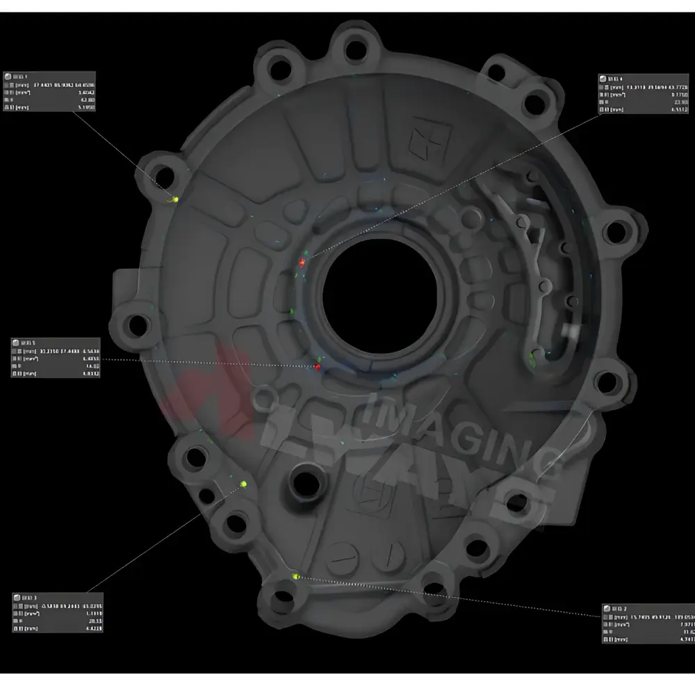
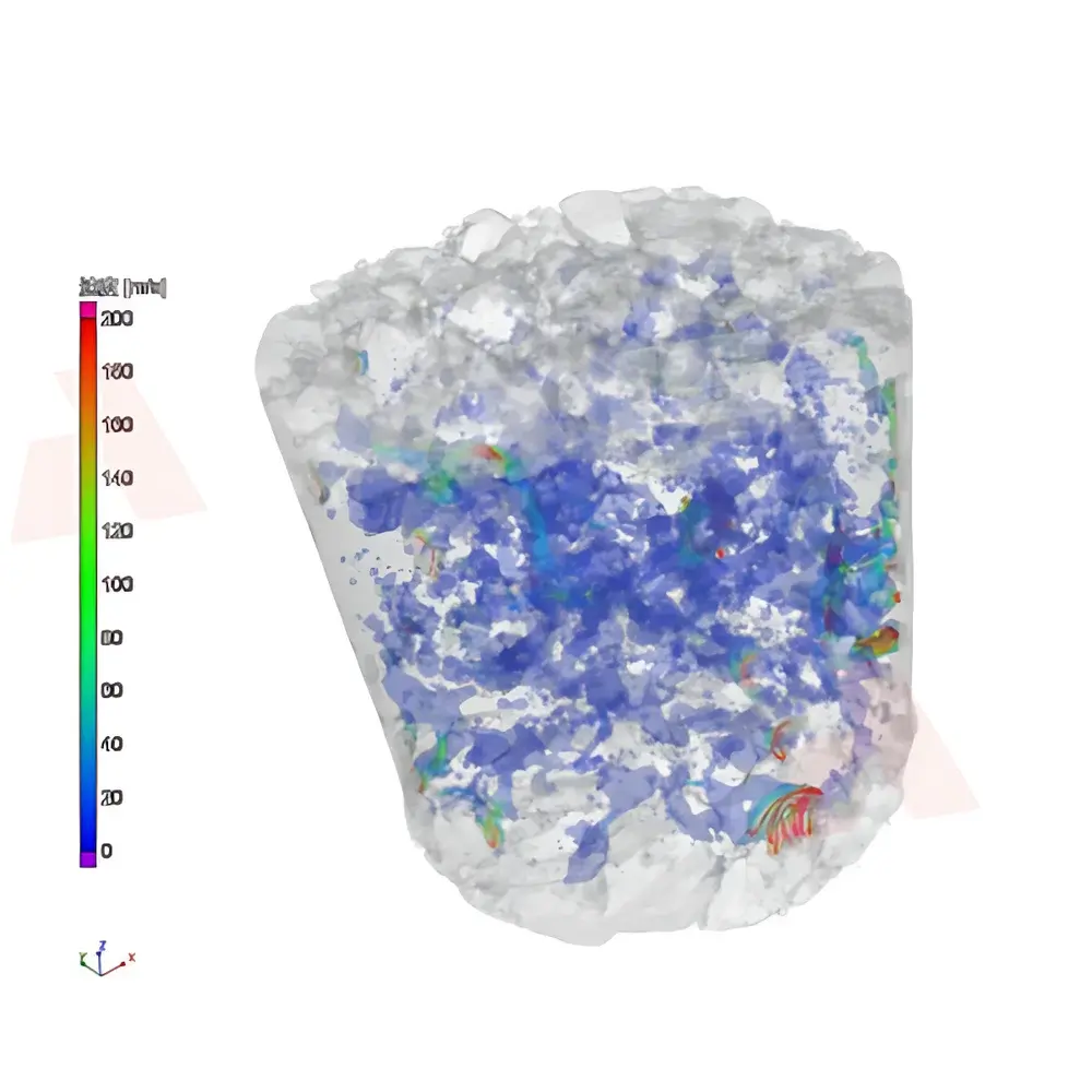
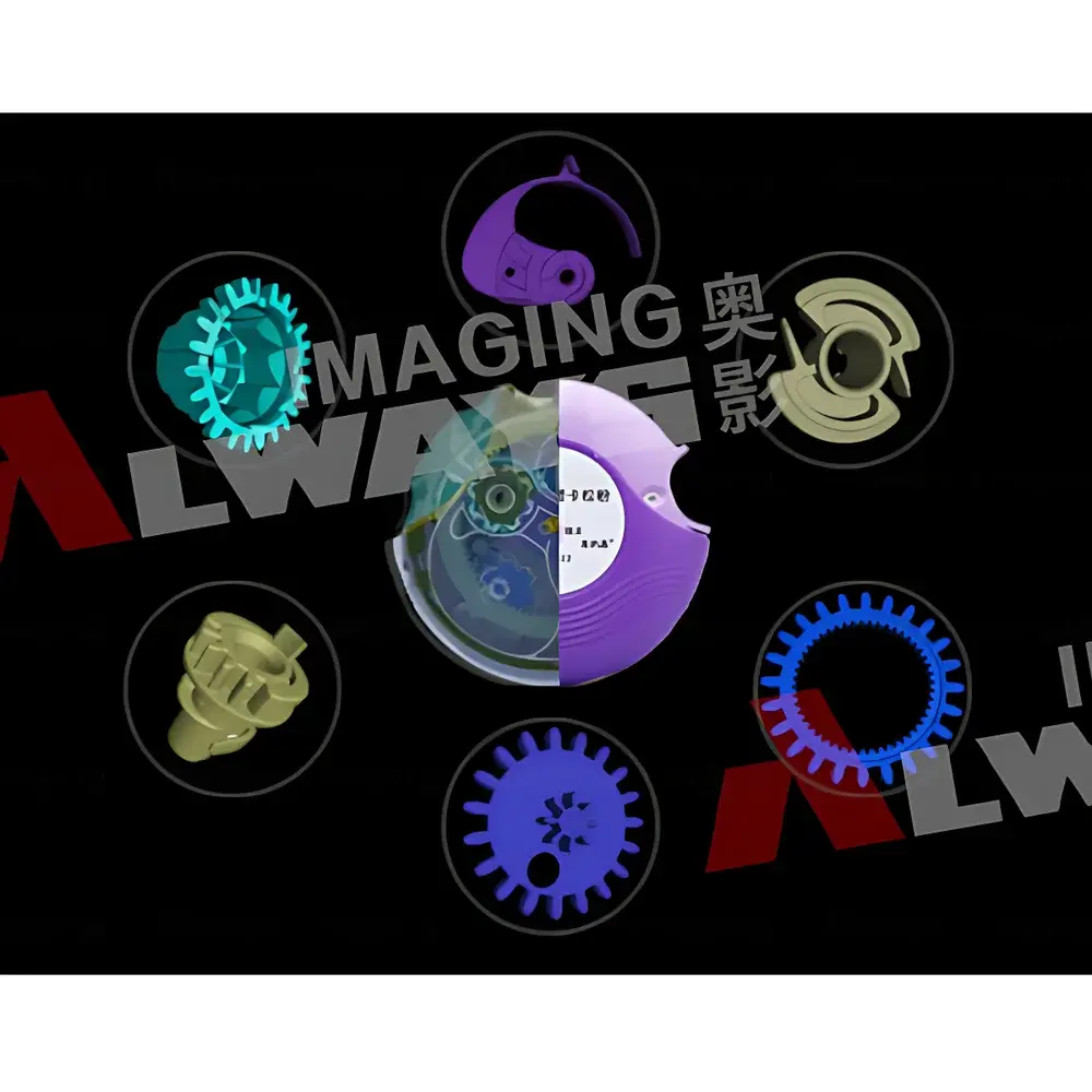
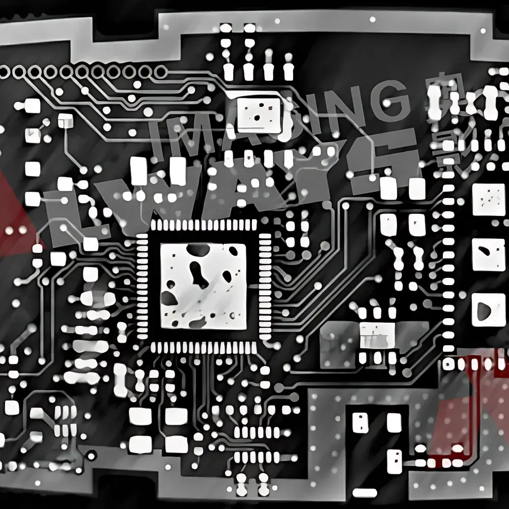
The AX-3000CT-D accommodates specimens up to Ø600 mm in diameter and 1000 mm in height, with maximum payload capacities of 50 kg, 100 kg, or 200 kg depending on configuration. It routinely images metallic castings (Al, Mg, Ti alloys), polymer composites, ceramic matrix materials, geological core samples (rock, soil, sediment), electronic assemblies (PCBs, solder joints, MEMS devices), biological tissues (fossilized bone, plant vasculature), and additively manufactured lattice structures. All system hardware and software comply with ISO 15708-1:2017 (Non-destructive testing — Qualification of computed tomography (CT) systems), ASTM E1441-21 (Standard Guide for Computed Tomography (CT) Imaging), and EU Directive 2013/59/Euratom for ionizing radiation protection. Optional audit-trail logging and user access control modules support GLP/GMP-aligned workflows per FDA 21 CFR Part 11 requirements.
Software & Data Management
Acquisition, reconstruction, and analysis are managed via Always Imaging’s proprietary CT Suite v5.x platform—a modular, scriptable environment built on CUDA-accelerated iterative reconstruction algorithms (SART, OS-SART, MBIR). The software supports DICOM-CT export, NRRD/HDF5 volumetric data interchange, and direct integration with third-party metrology tools (e.g., Volume Graphics VGStudio MAX, Thermo Fisher Avizo). Raw projection data is stored in lossless TIFF or HDF5 format with embedded metadata (source parameters, geometry calibration, exposure settings). Reconstruction pipelines include beam-hardening correction, ring artifact suppression, and phase-contrast enhancement (for low-Z materials). All processing steps are timestamped and logged with operator ID, supporting full traceability in regulated environments.
Applications
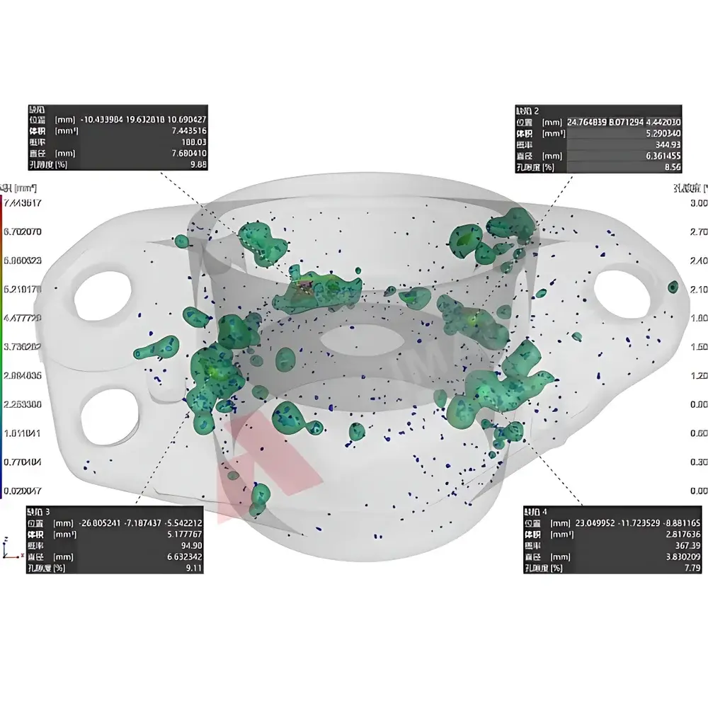
- Porosity and inclusion analysis in investment-cast aerospace turbine blades (ASTM E2905-20)
- Dimensional metrology of internal features in injection-molded medical device housings (VDI/VDE 2630-1.1)
- Microstructural characterization of battery electrode layers and solid-electrolyte interphases (SEI)
- Non-invasive paleontological examination of fossilized soft-tissue preservation and mineral replacement patterns
- Defect mapping in carbon-fiber-reinforced polymer (CFRP) laminates and adhesive bonds
- Quantitative analysis of pore network topology in reservoir rock cores for enhanced oil recovery modeling
- Failure analysis of hermetically sealed microelectronics under thermal cycling conditions
FAQ
What is the minimum resolvable feature size under optimal conditions?
Under nanofocus mode (180 kV, 0.5 mA, 1.5 µm focal spot) with high-magnification geometry and detector binning disabled, the system achieves an effective isotropic voxel size of 1.5–2 µm, validated using ISO 15708-3 line-pair phantoms.
Can the system perform dual-energy CT for material decomposition?
Yes—the dual-source architecture allows sequential or interleaved acquisition at two distinct kV settings (e.g., 180 kV + 240 kV), enabling basis-material decomposition and virtual monoenergetic reconstruction.
Is the system suitable for in situ mechanical testing?
With optional load frame integration (up to 50 kN axial capacity) and real-time reconstruction enabled, the AX-3000CT-D supports time-resolved 4D CT studies of deformation, crack propagation, and phase transformation under load.
How is geometric calibration maintained over time?
The system includes automated daily calibration routines using embedded reference spheres and edge targets; full volumetric calibration is performed annually using NIST-traceable step wedges and sphere arrays.
Does the software support automated defect detection and reporting?
Yes—CT Suite includes configurable AI-assisted segmentation models (U-Net architecture) trained on industrial defect libraries, with customizable pass/fail thresholds and ASME BPVC Section V-compliant reporting templates.


