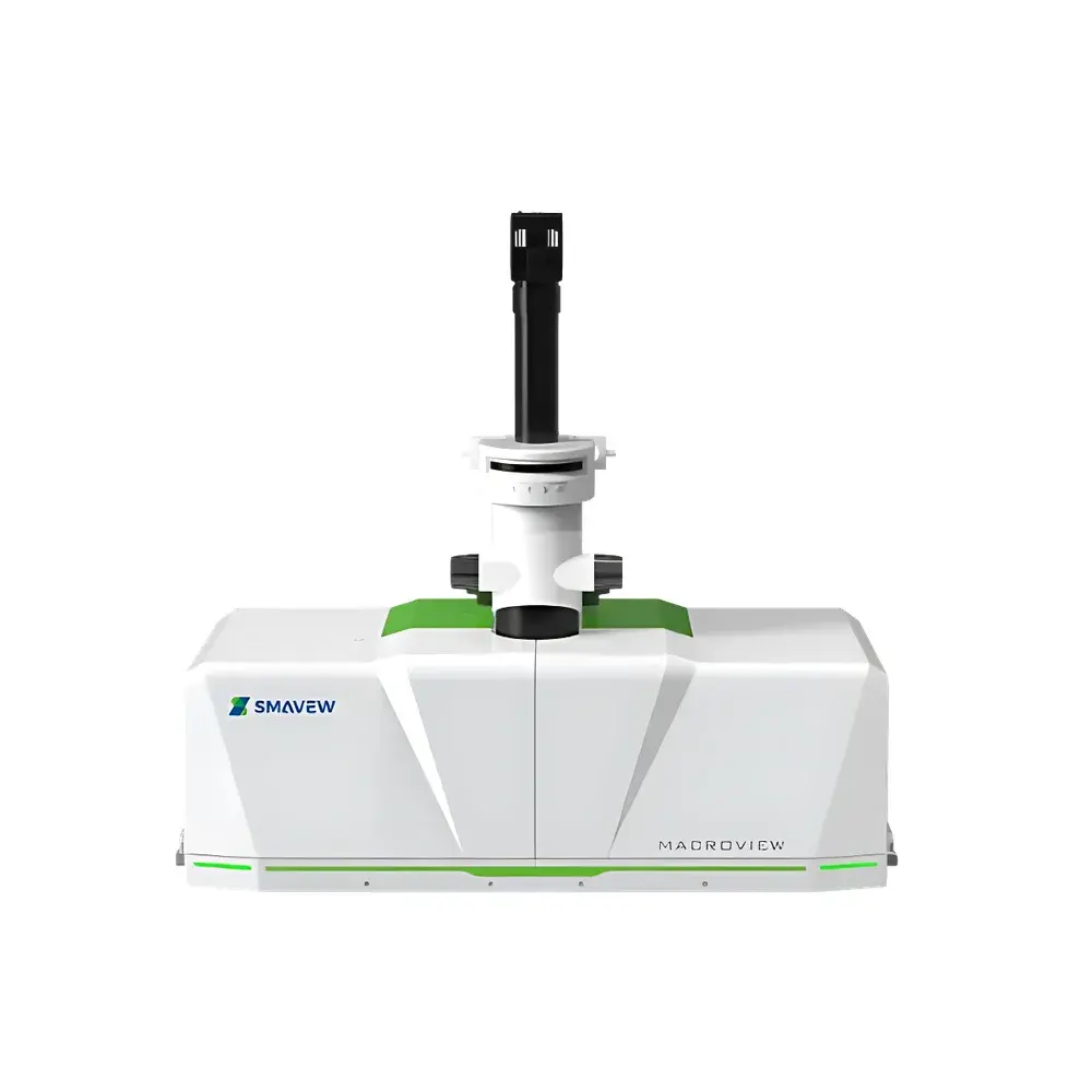
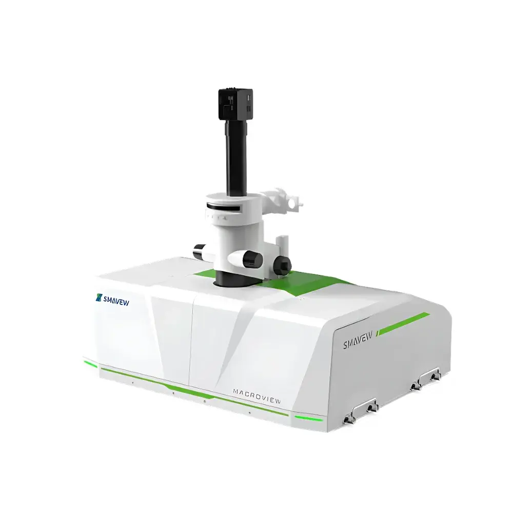
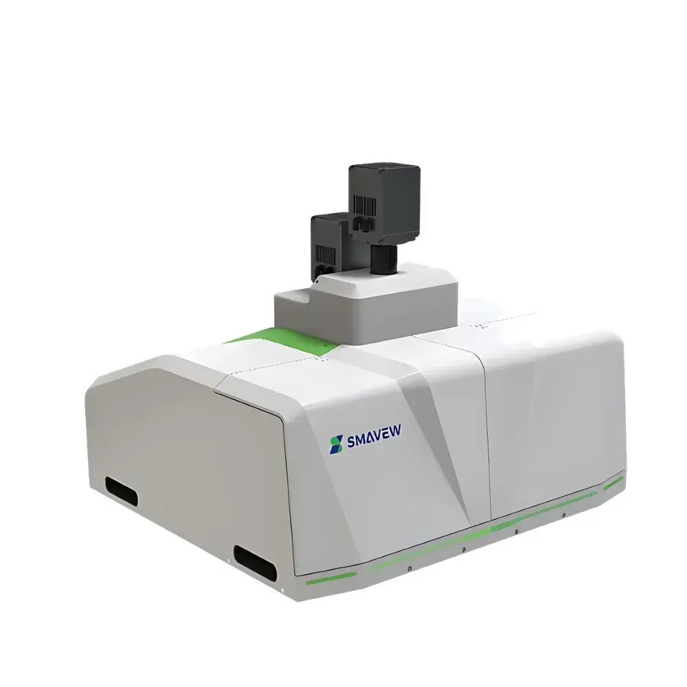
Smavew MacroView Ultra-High-Throughput Macro Light Sheet Microscope
| Brand | Smavew |
|---|---|
| Model | MacroView |
| Origin | Hubei, China |
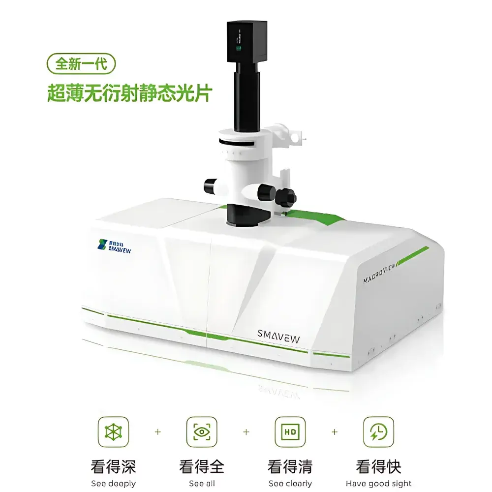
| Light Sheet Technology | Dr-SPIM (Diffractive-Robust Static Planar Illumination Microscopy) |
| Lateral Resolution | ≤300 nm |
| Axial Resolution | ≤1.5 µm |
| Sample Chamber Configurations | 5 independent chambers — two at 91 mm × 30 mm × 30 mm (organoids/tissues), two at 110 mm × 45 mm × 50 mm (whole organs, e.g., rat brain), one at 245 mm × 79 mm × 70 mm (whole mouse) |
| Stage Travel Range | ≥25 mm |
| Positioning Repeatability | ±≤1 µm |
| Laser Lines | 405 nm (violet diode), 488 nm (blue diode), 561 nm (green solid-state), 638 nm (red diode) |
| Objectives | Olympus 10×/0.6 (WD = 8 mm, RI = 1.33–1.52) |
| Camera | 2048 × 2048 pixels |
| Illumination | Dual-sided ultra-thin light sheet |
| Imaging Depth | Centimeter-scale |
| Data Acquisition Workstation | Intel Core i7-14700K or higher, ≥128 GB RAM, ≥1 TB OS SSD, dual 4 TB high-speed SSDs, 4 TB HDD, Windows 10 Pro 64-bit |
Overview
The Smavew MacroView Ultra-High-Throughput Macro Light Sheet Microscope is a purpose-built platform for volumetric fluorescence imaging of large, optically cleared biological specimens—from submillimeter organoids to intact adult mouse bodies. Engineered around the proprietary Dr-SPIM (Diffractive-Robust Static Planar Illumination Microscopy) optical architecture, MacroView generates stable, non-diffracting light sheets with sub-micron thickness control across centimeter-scale fields of view. Unlike conventional scanned or scanned-Bessel light sheet systems, Dr-SPIM employs static beam shaping and multi-parameter modulation to suppress axial sidelobes and minimize scattering-induced sheet distortion—enabling consistent optical sectioning fidelity over >10 mm axial depths without real-time adaptive correction. This architecture delivers isotropic resolution down to 1.5 µm axially and ≤300 nm laterally while maintaining high photon efficiency, thereby reducing phototoxicity and enabling rapid acquisition of terabyte-scale 3D datasets from whole-organ and whole-organism samples.
Key Features
- Dr-SPIM Optical Engine: Static, multi-modulated light sheet generation eliminates mechanical scanning artifacts and ensures uniform illumination intensity and thickness (≤1.5 µm) across full-field imaging volumes up to 245 mm in length.
- Modular Sample Chamber System: Five independently configurable chambers accommodate specimens spanning three orders of magnitude in volume: organoid-scale (91 × 30 × 30 mm), whole-organ (110 × 45 × 50 mm), and whole-mouse (245 × 79 × 70 mm), each optimized for refractive index matching and thermal stability during long-duration acquisitions.
- High-Fidelity Dual-Sided Illumination: Synchronized, co-aligned 405/488/561/638 nm laser paths on both sides of the sample enable balanced excitation and reduced shadowing—critical for quantitative multicolor imaging of heterogeneous tissues.
- Precision Motorized Stage: Linear stage with ≥25 mm travel range and ≤1 µm repeatability supports automated multi-position tiling and z-stack acquisition with sub-pixel registration accuracy.
- Optimized Objective Suite: Dual Olympus objectives—10×/0.6 (RI 1.33–1.52) for wide-field macro-imaging and 25×/1.0 (RI 1.41–1.52) for high-resolution subcellular detail—both featuring 8 mm working distance and chromatic aberration correction across the full laser spectrum.
Sample Compatibility & Compliance
MacroView is validated for use with standard tissue clearing protocols including CLARITY, CUBIC, iDISCO+, and SHIELD. Its chamber design accommodates aqueous, organic, and hybrid mounting media (e.g., FocusClear, RIMS, BABB) and supports continuous perfusion-compatible imaging for live-tissue preparations. The system complies with ISO 13485-aligned manufacturing controls and meets electromagnetic compatibility requirements per IEC 61326-1. All optical components are certified to RoHS and REACH standards. For regulated environments, the acquisition software supports audit trails, user access levels, and electronic signature functionality aligned with FDA 21 CFR Part 11 principles—facilitating GLP/GMP-compliant workflow documentation in preclinical research labs.
Software & Data Management
Acquisition and reconstruction are managed through Smavew’s MacroView Control Suite—a modular, Python-extendable platform supporting real-time deconvolution, adaptive background subtraction, and GPU-accelerated stitching of tiled volumes. The suite includes BIRDS (Brain Image Registration and Digital Segmentation), a dedicated module for Allen Mouse Brain Atlas v3 alignment. BIRDS performs nonlinear, pixel-level spatial registration of whole-brain datasets (up to 10 TB) using iterative mutual information optimization and hierarchical label propagation, enabling automated annotation of >1,000 neuroanatomical regions. Raw data export adheres to OME-TIFF and N5 formats; metadata conforms to the EMPIAR and BIDS-Imaging standards. Workstation configuration—Intel i7-14700K CPU, ≥128 GB DDR5 RAM, dual 4 TB NVMe SSDs, and 4 TB HDD—is rigorously tested for sustained throughput of ≥25 GB/min during multi-channel, multi-position acquisitions.
Applications
- Whole-organ 3D mapping of vascular, neuronal, and immune architectures in cleared human or rodent tissues.
- Rapid (<10 min) high-resolution whole-mouse brain imaging for connectomics and transgenic reporter quantification.
- Longitudinal developmental studies of embryonic organogenesis using light-sheet-based time-lapse of cleared embryos.
- Multi-scale phenotyping in disease models: e.g., tumor microenvironment visualization across intestinal segments, joint cartilage-bone interfaces, or spinal cord injury zones.
- Validation of spatial transcriptomics data via correlated 3D protein localization at organ-level resolution.
FAQ
What clearing methods are compatible with MacroView?
MacroView supports all major aqueous and organic clearing techniques—including CLARITY, CUBIC, uDISCO, and FRUIT—with chamber materials chemically inert to benzyl alcohol, dibenzyl ether, and ethyl cinnamate.
Can MacroView perform multi-color imaging with spectral unmixing?
Yes—the four-line laser combiner enables simultaneous or sequential excitation of common fluorophores (DAPI, FITC, TRITC, Cy5), and the software includes linear unmixing algorithms trained on reference emission spectra.
Is remote operation supported for core facility deployment?
The system supports secure remote monitoring and queue-based acquisition scheduling via TLS-encrypted web interface, compatible with institutional single-sign-on (SSO) infrastructure.
How is calibration traceability maintained?
Each instrument ships with NIST-traceable PSF beads (100 nm and 1 µm) and a digital calibration report documenting lateral/axial resolution, stage linearity, and laser power stability per channel.
Does MacroView support live-sample imaging?
While primarily designed for fixed, cleared specimens, optional environmental modules (temperature-controlled chamber, gas perfusion ports, and low-phototoxicity acquisition modes) enable limited live-tissue imaging of explanted organs or embryonic preparations.


