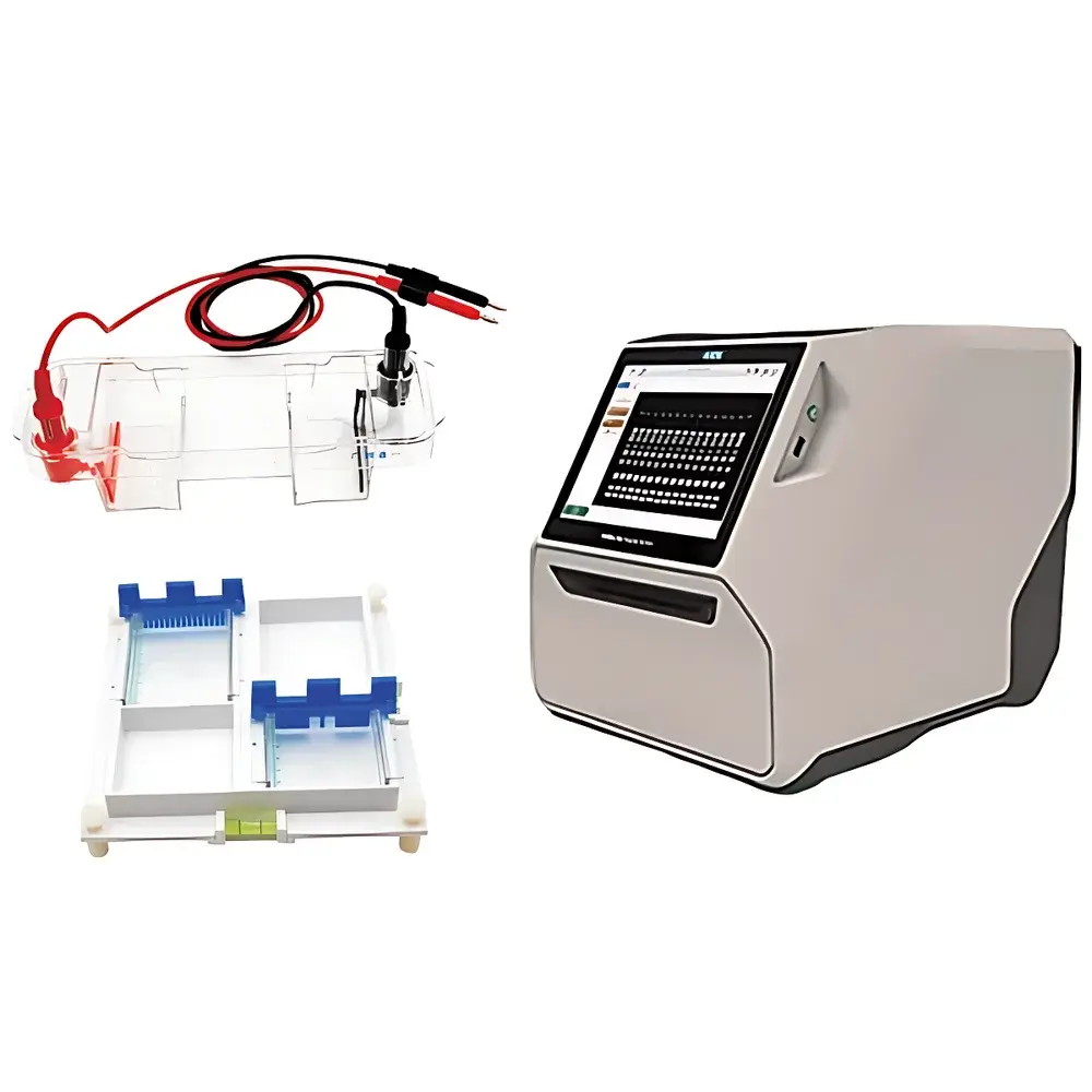
ACV NuacGel Series Nucleic Acid Gel Imaging System
| Brand | ACV |
|---|---|
| Origin | Shandong, China |
| Model | NuacGel Series |
| Instrument Type | Standard Gel Imaging System |
| Electrophoresis Module | Horizontal Benchtop Unit with Dual-Side Casting Platform |
| Imaging Module | Compact CMOS-Based UV/Visible Gel Documentation System |
| Power Supply | Programmable Constant Voltage/Constant Current Source with Real-Time Monitoring and Multi-Step Method Support |
| Compliance | Designed for ISO/IEC 17025-aligned laboratory workflows |
| Safety Features | Interlocked transparent chamber, UV shielded viewing window, emergency stop button, automatic power cutoff on lid opening |
| Sample Throughput | Up to four 70 mm × 70 mm or two 70 mm × 70 mm + two 70 mm × 100 mm gels simultaneously |
| Imaging Sensor | High-sensitivity monochrome CMOS sensor with adjustable exposure and auto-focus optics |
| Software | Image Lab Touch™ embedded interface with automated capture, background subtraction, band intensity quantification, molecular weight estimation, and export-ready TIFF/PNG/JPEG output |
Overview
The ACV NuacGel Series Nucleic Acid Gel Imaging System is an integrated horizontal electrophoresis and gel documentation platform engineered for reproducible, high-throughput nucleic acid analysis in academic, clinical, and quality control laboratories. It combines a precision-engineered horizontal electrophoresis unit—featuring dual-side gel casting capability, real-time visual monitoring through a fully transparent polycarbonate chamber, and programmable constant-voltage/constant-current power supply—with a compact, high-resolution imaging module optimized for ethidium bromide, SYBR® Safe, and other nucleic acid stains under UV (302 nm) and visible-light excitation. The system operates on the principle of electrophoretic separation in agarose or polyacrylamide matrices, followed by fluorescence detection and digital image acquisition. Its architecture supports routine applications including PCR product verification, restriction digest analysis, DNA ladder calibration, and plasmid purity assessment—all within a single, space-efficient footprint compliant with standard biosafety level 2 (BSL-2) lab infrastructure.
Key Features
- Dual-side gel casting platform enabling simultaneous preparation of up to four mini-gels (two 70 mm × 70 mm and two 70 mm × 100 mm formats) without reassembly or alignment recalibration
- Integrated bidirectional bubble-level indicators and four-point height-adjustable casting stand for sub-millimeter uniformity in gel thickness (±0.1 mm tolerance)
- Modular quick-release electrodes with color-coded connectors and physical alignment guides to eliminate insertion errors and simplify cleaning
- Interlocked transparent chamber with UV-filtering acrylic viewport and automatic power cutoff upon lid opening—meeting IEC 61010-1 electrical safety standards
- Programmable DC power supply supporting voltage (1–300 V), current (1–500 mA), and power (1–150 W) modes with real-time parameter logging and pause/resume functionality
- Onboard Image Lab Touch™ interface with touch-screen control, auto-exposure optimization, live preview, and one-click band quantification
- CMOS imaging sensor (effective resolution ≥ 5 megapixels) with adjustable gain, exposure time (0.1–60 s), and optical zoom for publication-grade image capture
Sample Compatibility & Compliance
The NuacGel system accommodates standard agarose (0.5–4%) and native/denaturing polyacrylamide (3–20%) gels. It is validated for use with common nucleic acid dyes including EtBr, SYBR® Gold, GelRed®, and GelGreen®. All hardware and firmware components are designed in accordance with ISO/IEC 17025:2017 general requirements for competence of testing and calibration laboratories. Data integrity features—including timestamped method storage, operator ID tagging, and immutable audit trails—support compliance with FDA 21 CFR Part 11 when used with validated software configurations. UV exposure during imaging is limited to user-defined durations and intensities, and optional UV shielding accessories meet ANSI Z87.1 impact and radiation protection criteria.
Software & Data Management
Image Lab Touch™ provides an embedded, intuitive operating environment with no external PC dependency. Functions include automatic background correction, lane and band detection using adaptive thresholding, relative quantification against internal standards, molecular weight interpolation from reference ladders, and customizable annotation layers. Export options include TIFF (uncompressed), PNG (lossless), and JPEG (optimized) formats, alongside CSV-formatted intensity and size data for downstream statistical analysis. Raw acquisition logs—including voltage, current, run duration, ambient temperature, and exposure settings—are stored in encrypted binary format with SHA-256 checksums to ensure traceability and prevent tampering.
Applications
- Verification of PCR amplification products and primer specificity
- Analysis of restriction enzyme digestion patterns for cloning validation
- Assessment of DNA fragmentation in apoptosis or shearing studies
- Quality control of plasmid preps and genomic DNA extractions
- Quantitative comparison of gene expression via RT-PCR band densitometry
- Educational demonstrations of electrophoretic mobility principles
FAQ
Can the NuacGel system be used for protein electrophoresis?
Yes—when paired with appropriate SDS-PAGE or native PAGE gels and compatible protein stains (e.g., Coomassie Blue, silver stain), though optimal sensitivity requires post-staining image enhancement protocols.
Is the power supply compatible with wet transfer applications?
Yes—the programmable electrophoresis module supports both standard gel electrophoresis and semi-dry/wet blot transfer protocols with selectable constant-current or constant-voltage modes.
Does the system support regulatory-compliant electronic signatures?
When deployed with validated networked software extensions and external identity management systems, full 21 CFR Part 11 compliance—including electronic signatures and role-based access control—is achievable.
What is the minimum detectable DNA mass under standard EtBr staining?
Under optimized conditions (100 ng loading, 302 nm UV excitation, 15 s exposure), detection limits range between 0.5–1.0 ng per band depending on fragment size and background uniformity.
Can third-party analysis software import NuacGel image files?
Yes—TIFF and CSV exports are natively supported by ImageJ/Fiji, Quantity One®, and Bio-Rad’s Image Lab desktop software without proprietary codec dependencies.
Related Products


