Makeway lgTIRF Fiber-Coupled Light-Guided Total Internal Reflection Fluorescence Microscopy System
| Brand | Makeway |
|---|---|
| Origin | Shanghai, China |
| Manufacturer Type | Authorized Distributor |
| Product Category | Domestic |
| Model | lgTIRF |
| Pricing | Upon Request |
| Measurement Principle | Light-guided TIRF (not SQUID) |
| Excitation Delivery | FC-PC fiber-coupled (2 m length) |
| Evanescent Field Depth | ~100 nm |
| Compatible Objectives | Dry, water-immersion, oil-immersion |
| Substrate Compatibility | Glass/silica coverslips, optically clear-bottom dishes |
| Excitation Wavelength Range | 190–1000 nm (UV-capable) |
| Configurable Modes | TIRF, SAFM (shallow-angle fluorescence), MSE (microspot excitation) |
| Calibration | Factory-calibrated, angle-stabilized geometry |
| Switching Speed | Instant mode switching between TIRF/SAFM/MSE/epifluorescence/transmission |
Overview
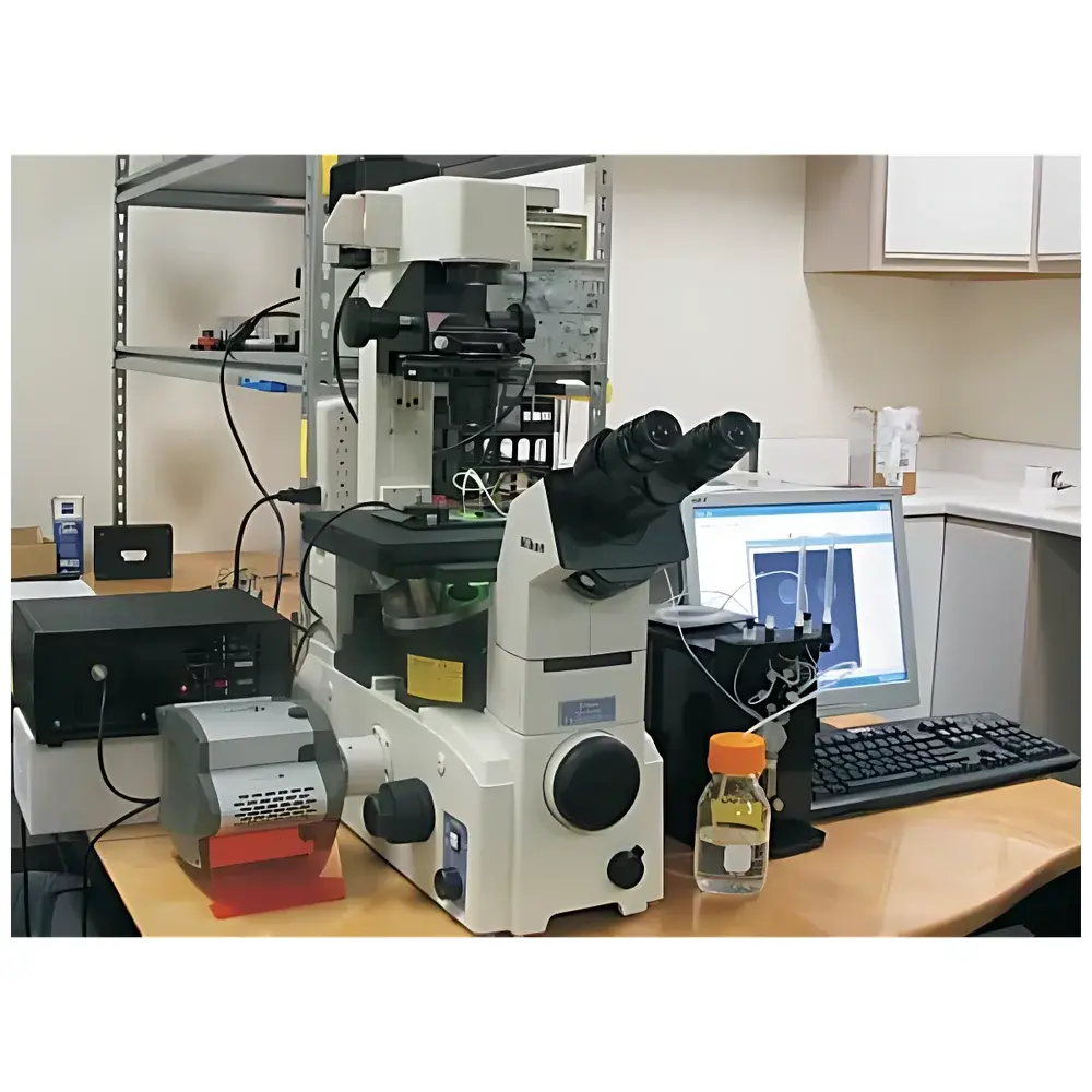
The Makeway lgTIRF is a fiber-coupled, light-guided total internal reflection fluorescence (TIRF) microscopy system engineered for high-fidelity evanescent field generation and quantitative single-molecule imaging. Unlike objective- or prism-based TIRF architectures, the lgTIRF decouples excitation from emission optics by delivering laser light via a 2-meter FC-PC terminated optical fiber to a precision-engineered silica light guide—typically a glass or fused-silica coverslip or optically clear-bottom culture dish. At the interface between the light guide and aqueous sample medium, total internal reflection occurs, generating an exponentially decaying evanescent wave with a characteristic penetration depth of approximately 100 nm. This physical constraint confines excitation exclusively to fluorophores within the immediate vicinity of the substrate surface, thereby suppressing out-of-focus background by >3–4 orders of magnitude relative to objective-TIRF systems. The system operates on a fixed-angle, factory-calibrated optical geometry, ensuring reproducible evanescent field intensity across repeated experiments and wavelength switches—eliminating realignment requirements. Its UV-transmissive fused-silica optical path (190–1000 nm) enables deep-UV excitation not feasible in conventional objective-TIRF setups, expanding compatibility with UV-sensitive probes and photoactivatable proteins.
Key Features
- Fiber-coupled architecture with 2 m FC-PC terminated silica fiber—enables flexible integration with multi-laser combiners, AOTFs, or monochromators
- Four interchangeable TIRF launch geometries: side-end coupling (SEL-1 narrow-band, SEL-7 wide-field), top-surface mobile coupler, and bottom-entry coupler for optically clear-bottom dishes
- Simultaneous support for dry, water-immersion, and oil-immersion objectives without optical reconfiguration
- Integrated shallow-angle fluorescence microscopy (SAFM) mode—excites fluorophores 1–5 µm above the surface using controlled low-angle incidence
- Microspot excitation (MSE) capability via tapered fiber probes (1–100 µm spot size), compatible with patch-clamp pipettes and dielectrophoretic manipulation
- UV-grade fused-silica optical components enabling excitation from 190 nm—critical for DAPI, Hoechst, and photoconversion applications
- Angle-stabilized, non-adjustable launch geometry ensures inter-experimental repeatability of evanescent field intensity
- Instant, software-controlled switching among TIRF, SAFM, MSE, epifluorescence, and transmission imaging modes
Sample Compatibility & Compliance
The lgTIRF system accommodates standard microscopy substrates including #1.5H glass coverslips (22 × 22 mm, 24 × 50 mm), fused-silica coverslips, and commercially available optically clear-bottom polystyrene or glass-bottom dishes (e.g., MatTek P35G-1.5-14-C, Ibidi µ-Dish 35 mm). It interfaces seamlessly with open perfusion chambers, sealed flow cells, and temperature-controlled stage-top incubators. All optical contact points—including fiber-to-coverslip coupling—are optimized for immersion oil (n = 1.518) to minimize Fresnel losses and maintain numerical aperture fidelity. The system conforms to ISO 10934-1 (optical microscopy terminology) and supports GLP-compliant experimental workflows through traceable calibration records and documented optical alignment protocols. While not itself a regulated medical device, its configuration adheres to design principles referenced in ASTM E2852-21 (standard guide for fluorescence microscopy validation) and supports data integrity practices aligned with FDA 21 CFR Part 11 when integrated with compliant acquisition software.
Software & Data Management
The lgTIRF operates as a hardware module under third-party acquisition platforms (e.g., Micro-Manager v2.0+, NIS-Elements AR, or MetaMorph), with full TTL and analog I/O control for synchronized laser gating, filter wheel positioning, and stage movement. Dedicated configuration files define each launch geometry’s effective incident angle and corresponding evanescent decay constant, enabling automated compensation during multi-wavelength acquisitions. All mode-switching events are timestamped and logged with metadata (wavelength, power, angle, substrate type), supporting audit trails required for method validation. Raw intensity profiles from calibration runs (e.g., fluorescent bead monolayers) can be exported in HDF5 or TIFF format for post-acquisition quantification of evanescent field uniformity (±3% spatial variation over 10 × 10 mm region). No proprietary closed-source software is required; all control logic resides in open API-accessible firmware.
Applications
The lgTIRF serves as a modular platform for surface-proximal fluorescence interrogation across multiple life science domains. In single-molecule biophysics, it enables high-contrast tracking of membrane-associated receptors (e.g., EGFR, integrins), synaptic vesicle docking kinetics, and real-time conformational dynamics of ion channels (e.g., TRPC family) using MSE-coupled patch pipettes. In cell biology, its compatibility with live-cell perfusion chambers facilitates long-term TIRF imaging of clathrin-mediated endocytosis, focal adhesion turnover, and lipid raft partitioning. For high-content screening, the SEL-7 wide-field emitter supports parallel readout of 20 × 20 mm microarrays—ideal for label-free binding assays and kinetic profiling of antibody-antigen interactions. SAFM mode extends utility to submembrane cytoskeletal dynamics (e.g., cortical actin flow) and organelle positioning studies where 1–5 µm axial resolution is optimal. Electrochemical and dielectrophoretic accessories allow simultaneous stimulation and imaging—enabling correlative electrophysiology-fluorescence experiments without optical compromise.
FAQ
Is the lgTIRF compatible with inverted and upright microscopes?
Yes—the system mounts on standard K-frame platforms (110 × 160 mm) and integrates with both inverted and upright configurations via adjustable XYZ translation stages.
Does lgTIRF require realignment when changing excitation wavelengths?
No—its fixed-angle light-guided geometry eliminates wavelength-dependent reoptimization; evanescent field depth scales predictably with λ per standard electromagnetic theory.
Can I use UV lasers (e.g., 266 nm) with this system?
Yes—UV-grade fused-silica optics and fiber enable stable transmission from 190 nm onward, unlike silica-doped objective lenses that absorb below 350 nm.
What is the typical evanescent field decay length, and how is it verified?
Measured decay lengths range from 70–120 nm depending on wavelength and refractive index mismatch; validated using supported lipid bilayer–anchored quantum dots and exponential fitting of axial intensity profiles.
How does lgTIRF compare to prism-based TIRF in signal-to-background ratio?
It achieves comparable SNR (>100:1 for single-molecule detection) while offering superior compatibility with open-chamber physiology rigs and multi-objective workflows.

")

