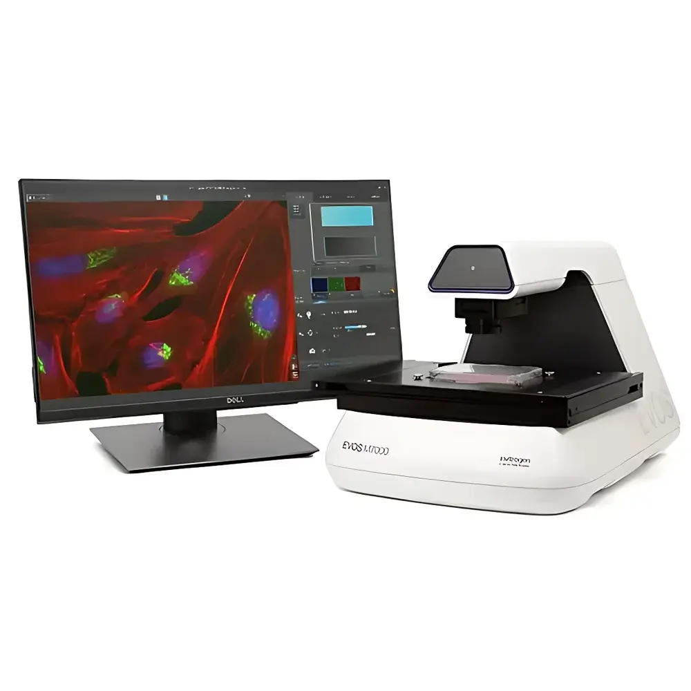
Thermo Fisher EVOS M7000 Intelligent Live-Cell Imaging and Analysis System
| Brand | Thermo Fisher |
|---|---|
| Origin | Shanghai, China |
| Manufacturer Type | Authorized Distributor |
| Product Category | Domestic (China-manufactured) |
| Model | EVOS M7000 |
| Pricing | Upon Request |
Overview
The Thermo Fisher EVOS M7000 Intelligent Live-Cell Imaging and Analysis System is a fully automated, inverted, multi-channel fluorescence and transmitted-light imaging platform engineered for high-fidelity, long-term live-cell observation under physiologically relevant conditions. Built upon an infinity-corrected optical architecture with 45 mm parfocal distance, the system employs LED-based illumination with hard-coated, precisely matched filter sets—eliminating mercury vapor lamps and enabling stable, low-phototoxicity excitation across four independent fluorescence channels plus brightfield, phase contrast, and color brightfield modes. Its core measurement principle relies on quantitative digital microscopy: spatially resolved pixel intensity acquisition from high-sensitivity CMOS sensors, synchronized with precise motorized stage motion, autofocus routines, and environmental control integration. Designed for reproducible high-content data generation in academic, pharmaceutical, and biotech laboratories, the EVOS M7000 supports time-lapse imaging, Z-stack acquisition, tiled mosaic scanning, and multi-position well-plate analysis—all programmable via an intuitive, touch-optimized interface without requiring pre-warm-up or cooling cycles.
Key Features
- Fully automated inverted microscope platform with motorized X/Y stage (120 mm × 80 mm travel, sub-micron resolution) and precision auto-focus mechanism
- 5-position objective turret compatible with a broad range of LWD and coverslip-corrected objectives (1.25× to 100×), including achromat, fluorite, and apochromat designs
- Dual high-sensitivity 3.2 MP CMOS cameras: monochrome sensor (2048 × 1536, 3.45 µm pixel pitch) optimized for fluorescence; color sensor for label-free or chromogenic applications
- Modular LED illumination system with >50,000-hour lifetime light cubes—each containing digitally controlled LEDs and hard-coated dichroics/beamsplitters for uniform, high-transmission excitation
- Integrated environmental control compatibility: seamless operation with optional EVOS Desktop Incubator for precise regulation of temperature (±0.2°C), O2 (1–21%), CO2, and humidity during extended time-lapse experiments
- 27-inch 4K display (3840 × 2160) and dedicated Dell XE9 workstation (Intel Core i7-11700K, 32 GB DDR4 RAM, 512 GB PCIe NVMe SSD, NVIDIA Quadro RTX4000 GPU) optimized for real-time image processing and large dataset handling
Sample Compatibility & Compliance
The EVOS M7000 accommodates diverse specimen formats—including glass slides, Petri dishes, culture flasks, and standard microplates (6–1536-well)—via interchangeable, lockable container holders with automatic alignment detection. Its long-working-distance condenser (60 mm, 4-position turret) and modular optical path support both plastic-bottom and glass-bottom vessels while maintaining optimal correction. The system complies with ISO 13485 design controls for medical device manufacturing environments and meets key regulatory expectations for GLP-compliant imaging workflows, including audit-trail-capable acquisition logs, user-access controls, and metadata-rich TIFF/OME-TIFF export. While labeled “For Research Use Only. Not for diagnostic procedures,” its hardware and software architecture aligns with foundational principles of FDA 21 CFR Part 11 for electronic records when deployed with validated Celleste Image Analysis software configurations.
Software & Data Management
The native EVOS Imaging Software provides a unified, workflow-centric interface supporting acquisition, visualization, and basic quantification. Key capabilities include Area View navigation (low-magnification overview with ROI definition), Field View (high-res single-field capture), Z-stack rendering, time-lapse movie generation (AVI/MP4), and automatic montage stitching across multiple fields. All acquisition parameters—including exposure time, LED intensity, focus offset, and stage coordinates—are saved as editable protocols with version history. Raw image data is stored in open-format TIFF with embedded OME metadata, ensuring interoperability with third-party analysis platforms. Optional Celleste Image Analysis Software extends functionality with AI-assisted segmentation, multi-parametric cell classification, kinetic profiling (intensity, area, shape, texture over time), and batch processing compliant with HCS-XML standards—enabling scalable, reproducible analysis for publication-grade datasets.
Applications
The EVOS M7000 serves as a primary imaging platform for quantitative cell biology studies requiring spatiotemporal fidelity. It is routinely deployed in neurobiology for neurite outgrowth tracking; immunology and immuno-oncology for co-culture dynamics and immune synapse formation; 3D spheroid and organoid imaging with Z-stack reconstruction; high-resolution IHC/IF quantification across tissue sections; and phenotypic screening in drug discovery pipelines. Its ability to maintain cells in controlled microenvironments during multi-day acquisitions makes it especially suited for assays involving metabolic stress response, apoptosis kinetics, mitotic progression, and stem cell differentiation—where phototoxicity minimization and signal stability are critical performance criteria.
FAQ
Is the EVOS M7000 compatible with existing EVOS accessories?
Yes—the system maintains full backward compatibility with all EVOS FL Auto hardware modules, including light cubes, objectives, container holders, and calibration tools.
Does the system support environmental control for hypoxic experiments?
Yes—when paired with the optional EVOS Desktop Incubator, the system enables programmable O2 control from 1% to 21%, along with temperature and humidity regulation, directly from the acquisition interface.
What file formats does the software export?
Native export includes OME-TIFF (with full metadata), TIFF, JPEG, PNG, AVI, and MP4; Celleste adds support for HCS-XML and CSV-based quantitative outputs.
Can the system perform automated multi-well plate imaging with Z-stacks?
Yes—fully programmable multi-position, multi-channel, multi-Z acquisition is supported across all standard microplate formats, with configurable dwell times and autofocus per well.
Is the LED illumination system field-serviceable?
LED light cubes are user-replaceable modules; no optical realignment is required upon installation due to factory-calibrated mechanical registration and beam homogenization.


