BeonChip BE-GRADIENT Electrochemical Gradient 3D Microfluidic Cell Culture Chip
| Brand | BEOnChip |
|---|---|
| Origin | Spain |
| Model | BE-GRADIENT |
| Chip Format | Standard microscope slide footprint (75 × 25 mm) |
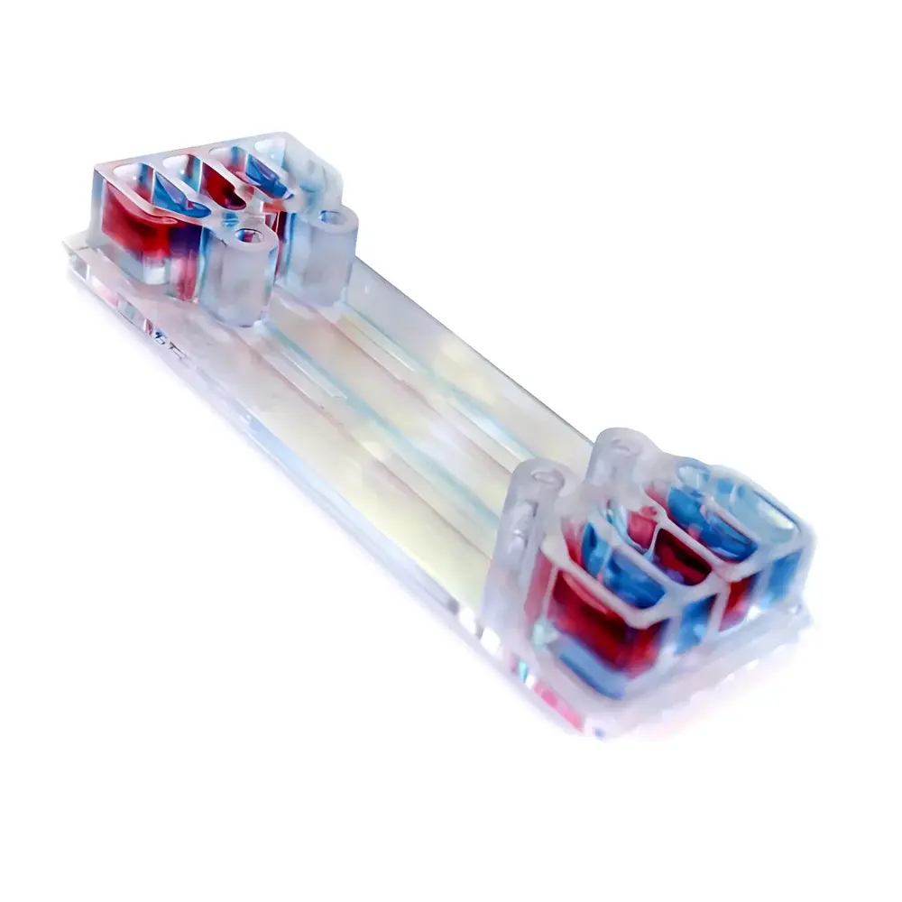
| Chamber Architecture | One central 3D culture chamber + three integrated microchannels (two lateral perfusion channels + one auxiliary channel) |
| Substrate Material | Optical-grade, plasma-bonded cyclic olefin copolymer (COC) |
| Transparency Range | 200–1100 nm (fully compatible with brightfield, phase contrast, DIC, and fluorescence microscopy) |
| Sterility | Pre-sterilized via ethylene oxide (EO) gas |
| Surface Treatment | Non-fouling, cell-adhesion-neutral coating (no extracellular matrix pre-coating required) |
| Fluidic Interface | Standard Luer-lock connectors (1/4"-28 thread) |
| Compatibility | Compatible with syringe pumps (e.g., Harvard Apparatus PHD Ultra), pressure-driven systems (e.g., Elveflow OB1), and automated perfusion platforms |
Overview
The BeonChip BE-GRADIENT is a purpose-engineered microfluidic chip designed for quantitative investigation of electrochemical and soluble gradient-driven cellular responses in physiologically relevant 3D microenvironments. Based on laminar flow physics and controlled diffusion principles, the device enables stable, reproducible establishment of linear or stepwise gradients—such as glucose, oxygen, chemokines, or redox-active species—across a central hydrogel-embedded cell culture region. Its architecture integrates a central gelation chamber (typical volume: ~2.5 µL) flanked by two parallel perfusion channels and one auxiliary channel, permitting independent control of inlet/outlet streams to generate bidirectional or asymmetric gradients. Unlike conventional Transwell or static gradient assays, the BE-GRADIENT sustains dynamic mass transport while preserving spatial resolution at single-cell level under continuous optical observation—making it suitable for time-lapse studies of directed migration, metabolic adaptation, and redox signaling.
Key Features
- Optically transparent COC substrate with <0.1% autofluorescence background across UV–NIR spectrum—validated for confocal and live-cell super-resolution imaging.
- Monolithic, single-use chip design eliminates assembly errors and cross-contamination; no bonding layers or glue interfaces that compromise gradient fidelity.
- Standardized microscope slide footprint (75 × 25 mm) ensures drop-in compatibility with inverted and upright widefield microscopes, motorized stages, and environmental chambers (37 °C / 5% CO₂).
- Non-adhesive surface chemistry prevents non-specific protein adsorption and enables recovery of intact 3D spheroids or organoids post-experiment via gentle enzymatic or mechanical dissociation.
- Luer-compatible fluidic ports support seamless integration with commercial syringe pumps, pressure controllers, and multi-channel perfusion manifolds—enabling programmable gradient ramping, pulsatile flow, or multi-analyte switching.
- Manufactured under ISO 13485-certified cleanroom conditions; EO-sterilized and individually packaged with certificate of conformance.
Sample Compatibility & Compliance
The BE-GRADIENT supports a broad range of primary and immortalized human and murine cell types—including endothelial cells, neurons, immune cells, and cancer lines—when encapsulated in collagen I, Matrigel®, fibrin, or synthetic PEG-based hydrogels. It has been validated for long-term (≥72 h) perfused culture with maintenance of physiological barrier integrity and metabolic activity. The chip complies with ISO 10993-5 (cytotoxicity) and ISO 10993-12 (sample preparation for biological testing). For GLP-regulated studies, full traceability documentation—including lot-specific sterility reports, material certificates, and dimensional inspection records—is available upon request. Device geometry and fluidic resistance profiles are consistent across production batches (CV < 3.2% for channel height, measured by profilometry).
Software & Data Management
While the BE-GRADIENT operates independently of proprietary software, its open-standard interface facilitates integration into automated workflows governed by third-party platforms such as LabVIEW, Python-based microfluidic control libraries (e.g., PyFluidics), or commercial systems supporting TTL/RS-232 protocols. Time-lapse image data acquired during gradient exposure can be annotated using FIJI/ImageJ with gradient vector mapping plugins. For regulatory submissions, raw image stacks, pump log files, and environmental sensor outputs may be archived in compliant formats (TIFF, CSV, HDF5) meeting FDA 21 CFR Part 11 requirements when paired with audit-trail-enabled acquisition software (e.g., NIS-Elements AR, ZEN Blue).
Applications
- Quantitative analysis of chemotactic and electrotactic cell migration under defined field strengths (0.1–5 V/cm) and ligand gradients (e.g., CXCL12, VEGF, SDF-1α).
- In vitro modeling of tumor angiogenesis and vascular co-culture dynamics under glucose or oxygen gradient stress.
- Investigation of metabolic zonation in hepatic or renal organoids exposed to opposing nutrient–waste gradients.
- Screening of neuroprotective compounds in neuronal-glial co-cultures subjected to oxidative stress gradients (e.g., H₂O₂, nitric oxide).
- Validation of biosensor performance in microphysiological systems where analyte diffusion kinetics must be decoupled from cellular consumption rates.
FAQ
Can the BE-GRADIENT be reused after sterilization?
No. It is a single-use, pre-sterilized device. Reuse compromises surface chemistry integrity and introduces risk of residual biomolecule carryover.
Is hydrogel polymerization possible directly inside the chip?
Yes. Temperature- and photo-initiated gelation (e.g., collagen at 37 °C, GelMA under 365 nm UV) is fully supported within the central chamber.
What minimum flow rate ensures stable gradient formation without shear-induced damage?
For most adherent cell types, laminar perfusion at 0.1–10 µL/min maintains Reynolds numbers < 0.1 and wall shear stresses < 0.5 dyn/cm²—within physiological ranges for microvascular endothelium.
Does BEOnChip provide protocol support for gradient calibration?
Yes. Application notes include fluorescent dye-based Peclet number validation methods (e.g., using FITC-dextran 40kDa) and computational fluid dynamics (CFD) boundary condition templates for COMSOL Multiphysics®.
Are custom channel geometries or material substitutions available?
Yes. BEOnChip offers OEM design services for modified aspect ratios, integrated electrodes, or alternative substrates (e.g., glass-PDMS hybrids) under NDA. Lead time: 8–12 weeks.


