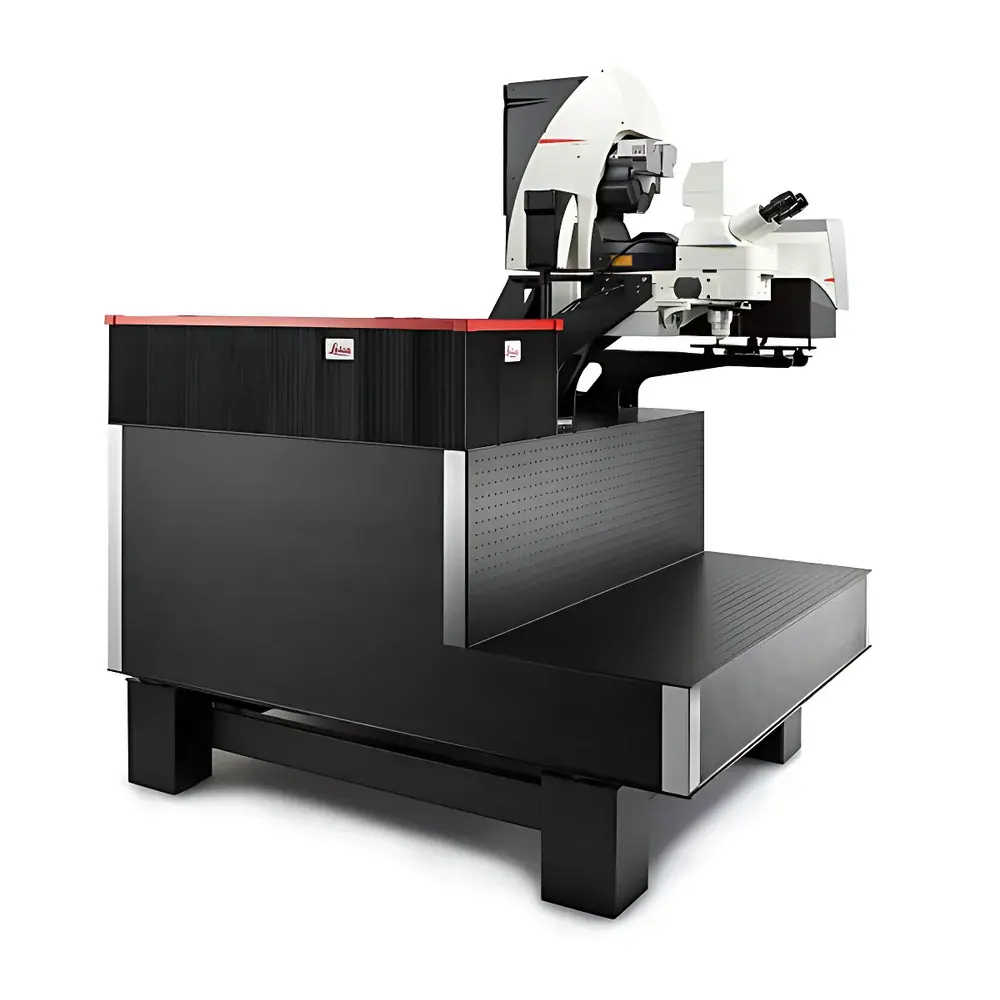
Leica STELLARIS 8 DIVE Spectral Multiphoton Microscope
| Brand | Leica |
|---|---|
| Origin | Germany |
| Model | STELLARIS 8 DIVE |
| Instrument Type | Point-Scanning Confocal Microscope |
| Excitation Range | Up to 1300 nm |
| Detection Range | 380–800 nm (tunable via 4Tune spectral detector) |
| Core Technology | Tunable non-descanned spectral detection with variable beam expander (VBE) |
| Compliance | Designed for GLP/GMP-aligned workflows |
Overview
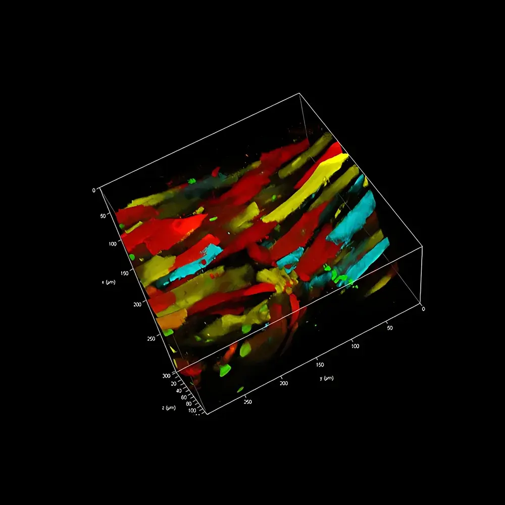
The Leica STELLARIS 8 DIVE (Deep In Vivo Explorer) is a high-performance spectral multiphoton microscope engineered for deep-tissue, multicolor in vivo imaging with unprecedented flexibility in spectral detection and optical path optimization. Built upon Leica’s proven point-scanning confocal architecture, the system integrates two foundational innovations: the patented 4Tune non-descanned spectral detection platform and the Variable Beam Expander (VBE). These technologies collectively address core limitations in conventional multiphoton microscopy—namely, fixed spectral bandpass constraints, chromatic aberration at infrared excitation wavelengths, and the inherent trade-off between imaging depth and lateral resolution. The STELLARIS 8 DIVE operates on the principle of nonlinear excitation (primarily two-photon and three-photon), where near-infrared (NIR) or infrared (IR) pulsed lasers induce fluorescence only at the focal plane, minimizing out-of-focus photodamage and enabling high-contrast volumetric imaging in scattering biological tissues up to several hundred micrometers deep. Its design prioritizes reproducibility, stability, and adaptability across evolving fluorescent probe landscapes—from genetically encoded biosensors (e.g., CFP, GFP, YFP, RFP) to synthetic dyes and lifetime-sensitive metabolic reporters (e.g., NADH, FAD, retinoids).
Key Features
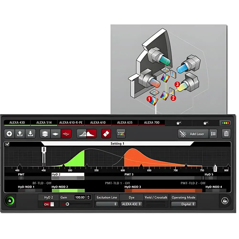
- 4Tune Spectral Detection System: A tunable, non-descanned detection architecture supporting 2–4 hybrid detectors (Power HyD NDDs and/or PMTs), enabling full-spectrum acquisition from 380 nm to 800 nm with user-defined spectral windows via drag-and-drop interface.
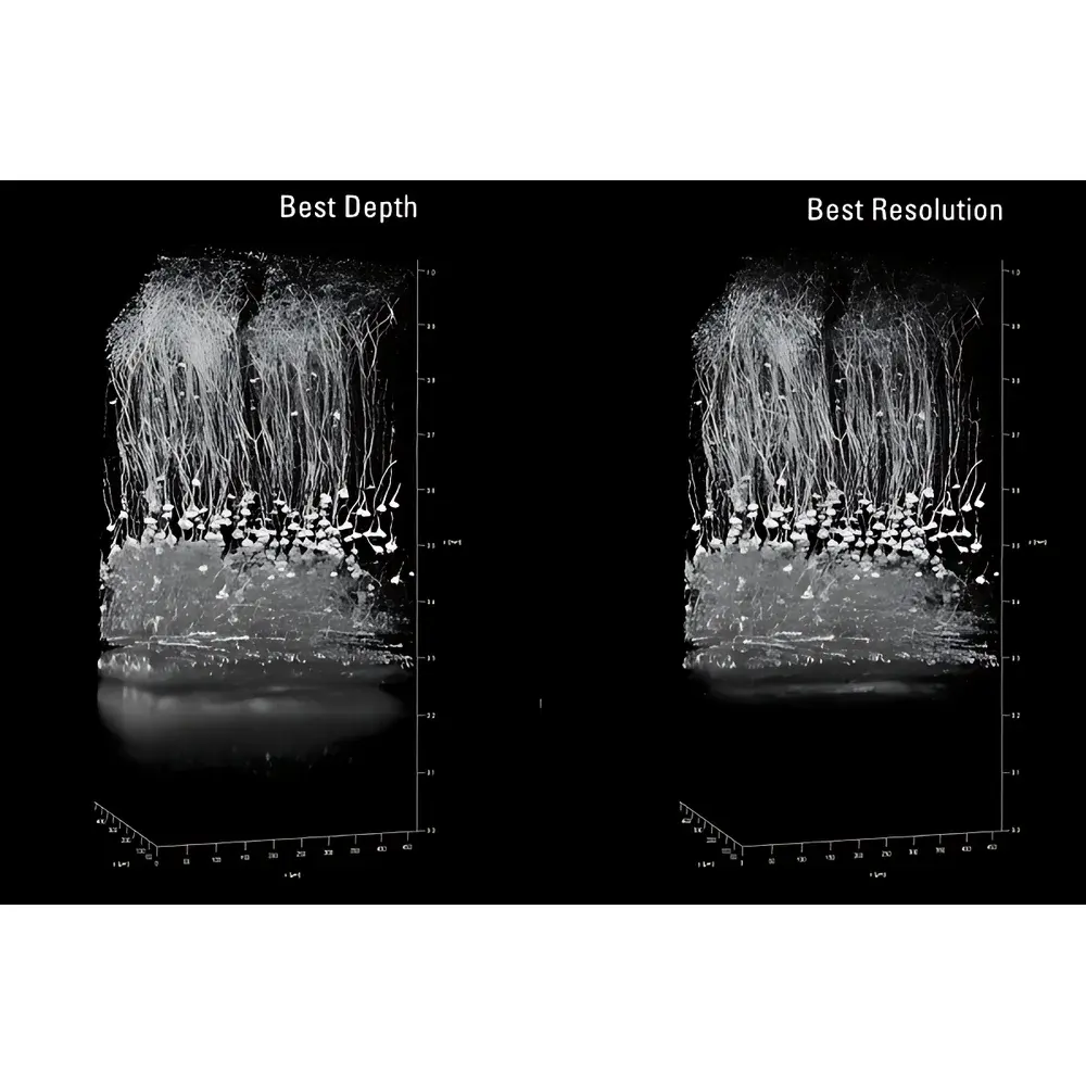
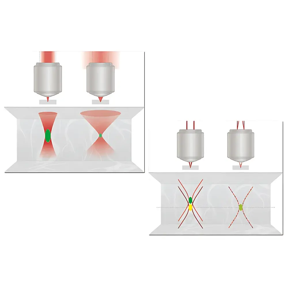
- Variable Beam Expander (VBE): Dynamically adjusts both beam diameter and divergence to balance resolution and penetration depth—optimal for IR-APO objectives (e.g., IRAPO 25×/1.0 W motCorr) and multi-wavelength excitation up to 1300 nm.
- Chromatic Aberration Correction: VBE compensates for longitudinal chromatic shift across visible and NIR bands, ensuring precise co-localization of multicolor signals without hardware realignment.
- Beam Stabilization & Overlap Recovery: Integrated beam collector actively maintains spatial overlap between visible and IR laser lines under thermal drift, mechanical vibration, or laser tuning—critical for quantitative in vivo co-localization studies.
- TauSense & FALCON Integration: Native compatibility with FLIM (Fluorescence Lifetime Imaging Microscopy) for metabolic phenotyping, redox state mapping, and unmixing of spectrally overlapping fluorophores based on lifetime signatures.
Sample Compatibility & Compliance
The STELLARIS 8 DIVE is validated for use with live animal models (e.g., murine cortex, zebrafish embryos, intestinal organoids), cleared tissue preparations (e.g., CLARITY, iDISCO), and thick 3D cell cultures. Its low-phototoxicity excitation strategy and adaptive detection make it suitable for longitudinal intravital imaging sessions exceeding 60 minutes. From a regulatory standpoint, the system—when deployed with Leica LAS X Navigator software—supports audit-trail logging, electronic signatures, and role-based access control aligned with FDA 21 CFR Part 11 requirements. It meets ISO 13485 design controls for research-use-only instrumentation and facilitates adherence to GLP principles in preclinical imaging studies. All optical components comply with IEC 60825-1:2014 Class 4 laser safety standards.
Software & Data Management
Control and analysis are unified within Leica LAS X software (v4.13+), which provides native support for spectral unmixing, lifetime fitting (FALCON), deconvolution, and 4D (x,y,z,t) data navigation. Raw spectral stacks are stored in standardized HDF5 format, ensuring interoperability with third-party analysis platforms (e.g., Python-based Napari, MATLAB, Imaris). The 4Tune UI enables real-time spectral window definition and batch reprocessing of previously acquired datasets—eliminating the need for repeat acquisitions when new fluorophores are introduced. Metadata—including laser power, dwell time, pinhole size, VBE settings, and detector gain—is embedded automatically and exportable in MIAME-compliant XML schema for reproducibility reporting.
Applications
- Longitudinal lineage tracing in intestinal crypts and neural stem cell niches using multicolor fluorescent reporters (CFP/GFP/YFP/RFP).
- Metabolic imaging of NADH/FAD redox ratios in awake, head-fixed mouse cortex during behavioral tasks.
- High-resolution structural mapping of amyloid plaques and microglial dynamics in Alzheimer’s disease models using far-red and NIR-II probes.
- Simultaneous optogenetic stimulation and functional calcium imaging with spatially confined photoactivation (<1 µm precision) enabled by multi-line excitation routing.
- Quantitative co-localization analysis across >4 spectral channels in cleared human tumor biopsies, corrected for depth-dependent chromatic shift via VBE calibration.
FAQ
What distinguishes STELLARIS 8 DIVE from conventional multiphoton systems?
It replaces fixed dichroic filters and PMT bandpasses with a fully tunable, non-descanned spectral detection engine (4Tune) and introduces active beam path optimization (VBE) to decouple resolution from penetration depth—enabling consistent sub-diffraction co-localization across >200 µm tissue depth.
Can the system be used for FLIM without hardware modification?
Yes. When equipped with FALCON-enabled detectors and synchronized PicoQuant or Toptica laser sources, the STELLARIS 8 DIVE performs time-resolved photon counting natively within LAS X software—no external TCSPC modules required.
Is the 4Tune detector compatible with third-party fluorophores?
Absolutely. Its continuous 380–800 nm spectral acquisition allows retrospective unmixing of any published fluorophore—even those developed after system installation—provided their emission spectra fall within the detection range.
How does the VBE improve imaging in scattering tissue?
By reducing effective numerical aperture (NA) at the objective back focal plane, the VBE elongates the focal volume along the axial direction while increasing photon density per voxel—enhancing signal-to-noise ratio and effective penetration without compromising lateral resolution.
Does Leica provide validation documentation for regulated environments?
Yes. Installation Qualification (IQ), Operational Qualification (OQ), and Performance Qualification (PQ) protocols are available upon request, including traceable calibration reports for laser power, spectral response, and stage positioning accuracy.


