Leica LMD Software for Laser Microdissection Systems
| Brand | Leica |
|---|---|
| Origin | Germany |
| Model | Leica LMD Software |
| Price Range | USD 13,500 – 27,000 (FOB Europe) |
| Regulatory Compliance | CE-marked, ISO 13485-aligned design controls, compatible with FDA 21 CFR Part 11–enabled audit trail configurations (when deployed with validated IT infrastructure) |
Overview
Leica LMD Software is the proprietary, integrated control platform engineered exclusively for Leica Microsystems’ Laser Microdissection (LMD) systems—including the Leica LMD6 and LMD7 platforms. Built on a deterministic real-time architecture, the software implements precise spatial coordination between high-resolution optical imaging, motorized stage positioning, and UV or IR laser beam delivery to enable contact-free, morphologically guided tissue or cell isolation at micron-level accuracy. Its core operational paradigm relies on pixel-accurate overlay of user-defined regions-of-interest (ROIs) onto live or stored microscopic images, followed by automated laser ablation along defined contours—ensuring reproducible sample harvesting without mechanical contact, thermal damage, or cross-contamination. Designed for routine use in translational research, clinical pathology, and single-cell omics workflows, the software supports both manual annotation and algorithm-assisted selection under multiple contrast modalities.
Key Features
- Fully integrated microscope control: native command layer for objective turret, focus drive, illumination intensity (transmitted/brightfield, fluorescence FLUO, differential interference contrast DIC, phase contrast PH, polarized light POL), and motorized XY/Z stage navigation
- Multi-modal ROI definition: vector-based drawing tools (freehand, polygon, rectangle, ellipse, Bézier curves) with adjustable anchor point density and sub-pixel edge refinement
- Three dedicated acquisition workflows: “Draw + Cut” for static tissue section excision; “Draw + Scan” for serial ablation across contiguous areas (e.g., slide-wide UV ablation for MALDI matrix deposition support); “Move + Cut” for dynamic real-time targeting during stage translation—optimized for touch-enabled displays
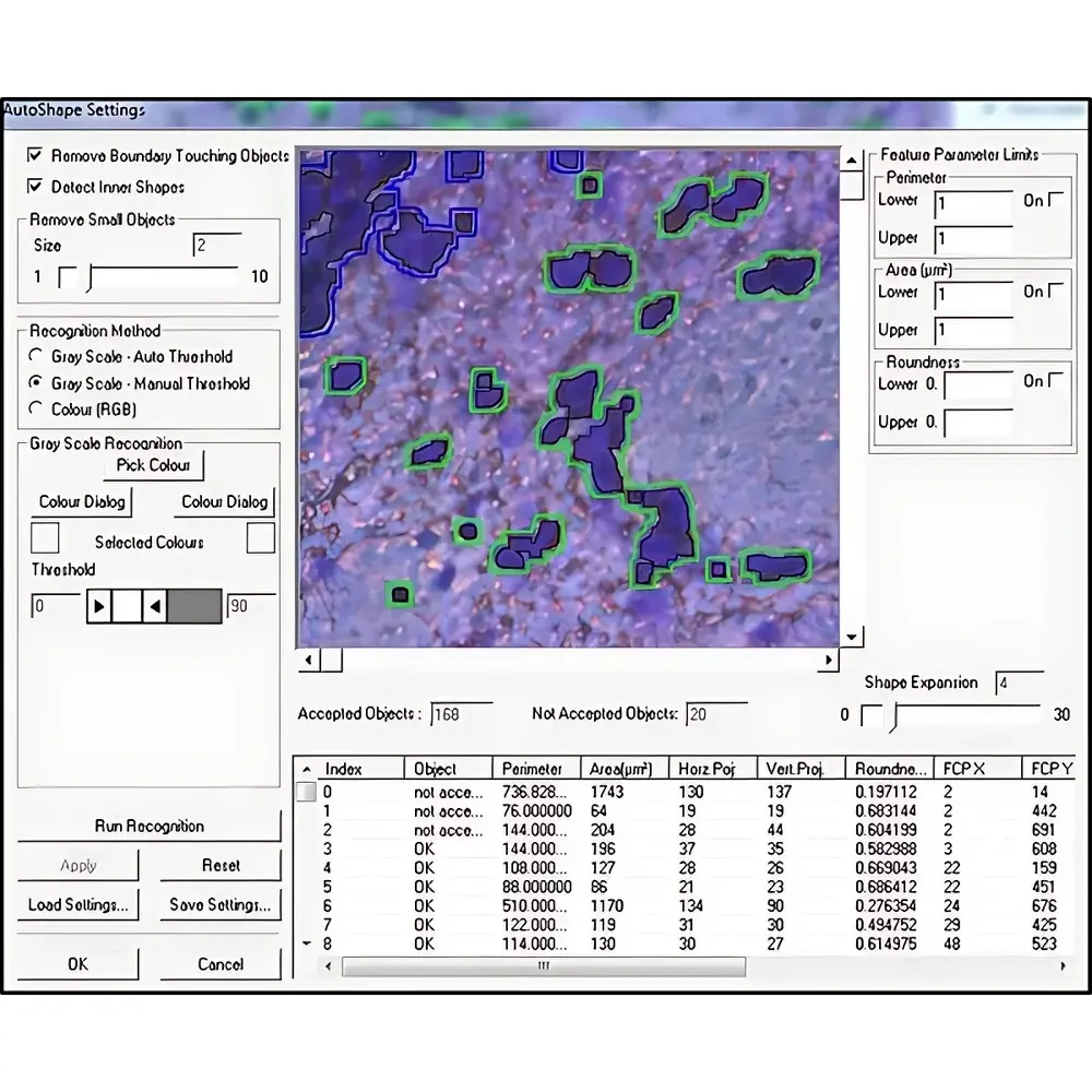
- Automated Cell Recognition (AVC) module: configurable machine learning–assisted segmentation using user-provided training sets; outputs binary masks for batch processing and metadata-tagged TIFF/OME-TIFF export
- Real-time preview rendering: GPU-accelerated image streaming from CMOS sensors with latency <120 ms; supports dual-display configuration for simultaneous navigation and ROI validation
- Audit-ready operation: optional electronic signature integration, time-stamped action logs, and configurable user permission tiers aligned with GLP/GMP documentation requirements
Sample Compatibility & Compliance
The software is validated for use with standard histological preparations including paraffin-embedded (FFPE) and frozen tissue sections (4–20 µm), cytospin preparations, and cultured monolayers on PEN-coated slides. It supports IHC- and IF-stained specimens, H&E, and unstained samples imaged via label-free contrast mechanisms. All system-level firmware and software updates undergo internal verification per ISO 14971 risk management principles. While the software itself is not a medical device, its deployment in diagnostic environments complies with EU IVDR Annex II essential requirements when used with CE-marked LMD hardware and documented SOPs. Export versions conform to EAR99 classification and include localized language packs (English, German, French, Japanese, Chinese).
Software & Data Management
Data handling follows FAIR principles: all acquired ROIs are stored as structured XML metadata alongside OME-TIFF image stacks containing channel-resolved pixel data, laser parameter history (pulse energy, repetition rate, dwell time), and stage coordinates. Batch export supports direct ingestion into downstream bioinformatics pipelines (e.g., Qiagen CLC Genomics Workbench, Partek Flow, or custom Python/R scripts via documented RESTful API endpoints). Local storage uses AES-256 encrypted SQLite databases; network deployment supports centralized license management and role-based access control via LDAP/Active Directory integration. Version history is maintained across major releases (v4.x → v5.x), with backward compatibility guaranteed for project files generated within the prior two major versions.
Applications
- Single-cell and microregion transcriptomics (e.g., spatial RNA-seq library prep from defined cortical layers)
- Proteomic profiling of tumor subclones isolated from heterogeneous FFPE blocks
- Microbial colony picking from agarose-embedded environmental samples
- Contamination-free isolation of meiotic cells for karyotype analysis
- Preparation of homogeneous cell populations for CRISPR screening validation
- Forensic trace evidence micro-extraction (e.g., spermatozoa from epithelial background)
FAQ
Is Leica LMD Software compatible with third-party microscopes?
No. It is licensed and functionally locked to Leica LMD-series hardware platforms only. Interfacing with non-Leica optics requires custom middleware development not supported under warranty.
Does the AVC module require separate licensing?
Yes. Automated Cell Recognition is an optional add-on module requiring annual maintenance subscription and separate activation key.
Can cutting parameters be saved and recalled per project type?
Yes. Users define and store parameter presets—including laser wavelength, pulse duration, scanning speed, and focus offset—for specific tissue types or staining protocols. Presets are embedded in project templates.
What file formats does the software natively export?
OME-TIFF (with multi-channel and Z-stack support), XML (ROI geometry and acquisition metadata), CSV (stage coordinates and timestamps), and PDF (annotated report summaries).
Is remote operation supported over LAN/WAN?
Local network control is fully supported; wide-area remote desktop access is permitted but not officially validated for GxP environments due to uncontrolled network latency and encryption boundary risks.


