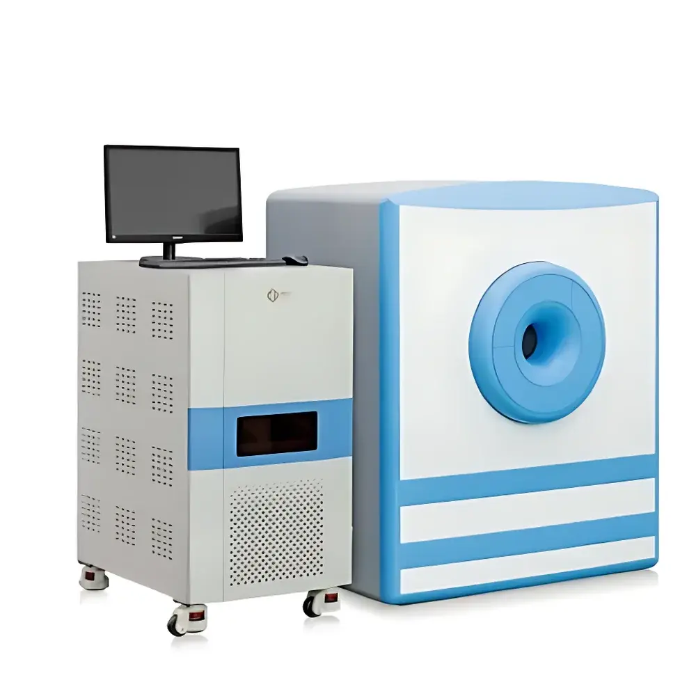
NIUMAG NM42-060H-I Small Animal MRI System
| Brand | NIUMAG |
|---|---|
| Origin | Jiangsu, China |
| Manufacturer Type | Authorized Distributor |
| Country of Origin | China |
| Model | NM42-060H-I |
| Magnetic Field Strength | 1.0 ± 0.05 T |
| Larmor Frequency | ~42 MHz |
| Gradient Coil Bore Diameter | 60 mm |
| Magnet Type | Permanent Magnet |
| Application Scope | In vivo small animal MRI (mouse/rat), contrast agent evaluation, tumor monitoring (brain, subcutaneous, liver), obesity phenotyping, longitudinal therapeutic response assessment |
| Compliance | Designed for GLP-aligned preclinical research environments |
Overview
The NIUMAG NM42-060H-I is a dedicated permanent-magnet-based small animal magnetic resonance imaging (MRI) system engineered for high-contrast, non-invasive in vivo visualization of soft tissue anatomy and pathophysiology in murine models. Operating at a nominal static magnetic field strength of 1.0 ± 0.05 T—corresponding to a proton Larmor frequency of approximately 42 MHz—the system leverages robust spin-echo and gradient-echo pulse sequences to generate quantitative T1-, T2-, and PD-weighted images with inherent soft-tissue differentiation. Unlike cryogen-dependent superconducting systems, its permanent magnet architecture eliminates helium dependency, reducing infrastructure requirements and long-term operational overhead while maintaining field homogeneity sufficient for reproducible longitudinal studies. The 60 mm horizontal bore accommodates standard mouse and juvenile rat positioning, supporting both anesthetized and restrained physiological imaging protocols under controlled environmental conditions.
Key Features
- Permanent magnet platform delivering stable 1.0 T field with <0.1 ppm/h temporal drift and <10 ppm spatial homogeneity over a 30 mm DSV (Diameter Spherical Volume)
- Dedicated 60 mm inner-bore gradient coil optimized for high-resolution anatomical imaging in mice (typical in-plane resolution: 100–200 µm; slice thickness: 0.5–1.0 mm)
- Integrated RF transceiver with tunable quadrature volume coil (60 mm ID) supporting proton (¹H) detection only—no multinuclear capability
- Real-time shimming via automated first-order shim routines prior to each acquisition sequence
- Modular software architecture enabling protocol customization, DICOM export, and batch processing of multi-timepoint datasets
- Low-power consumption design (<3 kW total system draw), compatible with standard laboratory AC power supply (220 V, 50 Hz)
Sample Compatibility & Compliance
The NM42-060H-I is validated for use with C57BL/6, BALB/c, nude, and NSG mouse strains across weight ranges of 18–35 g, as well as postnatal day 14–21 rats (up to 60 g). Anesthesia integration supports isoflurane delivery via calibrated vaporizer and MR-compatible respiratory gating hardware. All imaging protocols adhere to the ARRIVE 2.0 guidelines for reporting animal research. While the system itself does not carry FDA 510(k) or CE-IVD certification, its output data meets the structural and metadata requirements for inclusion in GLP-compliant preclinical study reports per OECD Test Guidelines 407 and 422. Image metadata conforms to DICOM 3.0 Part 10 standards—including modality (MR), manufacturer (NIUMAG), model name (NM42-060H-I), and acquisition parameters—for seamless integration into PACS or ELN platforms.
Software & Data Management
Acquisition and reconstruction are managed through NIUMAG’s proprietary MICE-Scan v4.x suite, built on a Qt-based GUI with command-line scripting support (Python API available). Core functionalities include: interactive ROI definition with intensity histogram analysis; automated brain segmentation using atlas-matched registration (Allen Mouse Brain Common Coordinate Framework v3); T2 mapping via monoexponential curve fitting of multi-echo spin-echo series; and dynamic contrast-enhanced (DCE) MRI parameter extraction (Ktrans, ve) using Tofts modeling. Raw k-space data is stored in vendor-neutral NIfTI-1 format; processed images export to DICOM, PNG, TIFF, or MATLAB .mat files. Audit trails log user login, sequence execution, parameter modification, and export events—supporting traceability requirements under ISO/IEC 17025 and internal QA procedures.
Applications
- Tumor phenotyping: Longitudinal tracking of orthotopic glioblastoma (GL261), subcutaneous melanoma (B16-F10), and hepatic metastasis models with volumetric quantification and necrosis characterization
- Metabolic disease modeling: Quantitative fat fraction mapping in liver and visceral adipose tissue using chemical shift–encoded MRI (Dixon technique)
- Neuroinflammation assessment: Contrast-enhanced BBB permeability analysis following LPS challenge or EAE induction
- Cardiac functional analysis: Cine-MRI for left ventricular ejection fraction (LVEF), stroke volume, and wall motion scoring in pressure-overload models
- Contrast agent development: In vivo relaxivity (r₁, r₂) determination and biodistribution kinetics of Gd- or Mn-based agents
FAQ
Is the NM42-060H-I suitable for functional MRI (fMRI) studies?
No—this system lacks the gradient slew rate, temporal resolution, and EPI-capable sequence library required for BOLD fMRI. It is optimized for structural and quantitative relaxation mapping.
Does the system support multi-nuclear imaging (e.g., ³¹P, ¹⁹F)?
No—hardware and software are configured exclusively for ¹H detection. No broadband RF amplifier or frequency-synthesizer tuning range is provided for alternate nuclei.
What level of technical support is available outside mainland China?
NIUMAG provides remote diagnostics, protocol optimization consulting, and software updates via secure VPN; on-site service requires coordination with authorized regional partners under contractual SLA terms.
Can raw k-space data be exported for third-party reconstruction?
Yes—unprocessed k-space matrices (complex float32) are accessible via MICE-Scan’s “Export Raw K-Space” module and compatible with MATLAB, Python (SciPy, SigPy), or ISMRM-recommended reconstruction pipelines.
Is the magnet shielded to reduce fringe field exposure?
Yes—the permanent magnet assembly includes passive ferromagnetic shielding, limiting the 0.5 mT contour to within 1.2 m from the bore center—compliant with ICNIRP occupational exposure limits for static fields.


