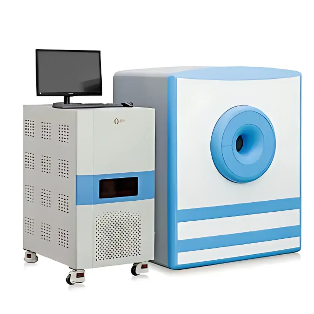
NIUMAG NM42-0 Small Animal MRI System
| Brand | NIUMAG |
|---|---|
| Origin | Jiangsu, China |
| Model | NM42-0 |
| Field Strength | 1.0 T Permanent Magnet |
| Maximum Sample Width | <40 mm |
| Animal Weight Range | 1–45 g |
| Minimum Sample Volume | ≥100 µL |
| Minimum Slice Thickness | 0.8 mm |
| Imaging Modality | Nuclear Magnetic Resonance (NMR)-based Magnetic Resonance Imaging (MRI) |
| Regulatory Classification | Preclinical In Vivo Imaging System |
| Compliance Framework | Designed for GLP-compliant preclinical research environments |
| Software | Integrated MRI acquisition platform with optional advanced image processing module |
Overview
The NIUMAG NM42-0 Small Animal MRI System is a compact, high-stability 1.0 Tesla permanent magnet-based magnetic resonance imaging platform engineered specifically for non-invasive, longitudinal in vivo studies in preclinical rodent models. Unlike X-ray or optical modalities, this system leverages nuclear magnetic resonance physics—detecting proton spin relaxation behavior in biological tissues under controlled static and gradient magnetic fields—to generate quantitative, three-dimensional anatomical and functional contrast without ionizing radiation. Its homogeneous 1.0 T field enables high signal-to-noise ratio (SNR) acquisition and spatial resolution sufficient for sub-millimeter structural delineation (down to 0.8 mm slice thickness), supporting reproducible morphometric analysis across repeated timepoints. The system is purpose-built for laboratory environments lacking dedicated RF-shielded rooms, eliminating infrastructure barriers commonly associated with superconducting MRI platforms.
Key Features
- 1.0 T permanent magnet architecture delivering stable, drift-free field homogeneity (<5 ppm over 30 mm DSV), enabling robust T1/T2 mapping and high-fidelity anatomical imaging.
- Non-ionizing, non-invasive operation—no radiation exposure, no tissue stress, fully compatible with longitudinal study designs in live mice (1–45 g) and other small mammals with transverse dimension <40 mm.
- Streamlined workflow: automated parameter optimization reduces operator dependency; full MR acquisition achievable in three intuitive interface steps without requiring advanced NMR theory knowledge.
- Modular hardware integration: supports optional gas anesthesia delivery systems (isoflurane/O₂), physiological monitoring interfaces (respiratory gating, temperature control), and RF coil adaptors for targeted anatomy (e.g., brain, abdomen, extremity).
- Low total cost of ownership: no cryogens, no RF shielding room requirement, minimal routine maintenance, and zero consumables beyond standard animal handling supplies.
- Open pulse sequence architecture: users may adjust RF pulse width, amplitude, repetition time (TR), echo time (TE), and trigger delays—facilitating method development for relaxometry, diffusion-weighted imaging, or custom contrast agent validation protocols.
Sample Compatibility & Compliance
The NM42-0 accommodates a broad range of preclinical sample types: intact anesthetized mice and rats (≤45 g); excised organs or tissue specimens placed in standardized NMR tubes; aqueous suspensions including nanoparticle colloids, ionic solutions, and microbial cultures (minimum volume: 100 µL). All acquisitions adhere to ISO/IEC 17025-aligned instrument performance verification procedures. While not FDA-cleared for human use, the system meets essential design criteria referenced in ASTM E2915-21 (Standard Practice for Evaluating Preclinical MRI Systems) and supports data integrity practices aligned with GLP and 21 CFR Part 11 when paired with audit-trail-enabled software configurations. Image metadata includes DICOM-compliant headers (including manufacturer, model, field strength, sequence parameters, and acquisition timestamps) for traceability in regulatory submissions.
Software & Data Management
The integrated MRI acquisition software provides real-time preview, sequence selection (Spin Echo, Gradient Echo, Inversion Recovery, Multi-Slice 2D/3D), and on-the-fly parameter tuning via graphical sliders. It logs all acquisition settings, hardware states, and environmental conditions (coil temperature, magnet stability metrics) to structured CSV and XML files. The optional Advanced Image Processing Suite operates as a standalone DICOM viewer and analysis engine, supporting ROI-based quantification (T1/T2 relaxation times, signal intensity ratios), pseudo-color mapping, multi-planar reformatting (MPR), 3D surface rendering, distance/angle measurement tools, threshold segmentation, and batch export to MATLAB, Python (NumPy), or Excel-compatible formats. All processed datasets retain original DICOM provenance tags to ensure chain-of-custody compliance.
Applications
- Contrast agent development: quantitative r1/r2 relaxivity assessment in vitro and in vivo using standardized phantoms and tumor-bearing murine models.
- Oncology research: longitudinal monitoring of orthotopic or subcutaneous tumor volume, necrosis fraction, and treatment-induced changes in perfusion or cellularity.
- Neuroscience: high-resolution structural imaging of mouse brain anatomy, hippocampal volumetry, and white matter tractography feasibility studies.
- Nanomaterial biodistribution: tracking iron oxide or gadolinium-based nanoparticles via T2* signal void quantification in liver, spleen, and tumor microenvironments.
- Microbiology: rapid concentration estimation of bacterial or yeast suspensions through transverse relaxation rate (R2) correlation with optical density or CFU counts.
- Pharmacokinetic modeling: dynamic contrast-enhanced (DCE) MRI for estimating vascular permeability (Ktrans) and extracellular-extravascular space (ve) in disease models.
FAQ
Does the NM42-0 require a dedicated RF-shielded room?
No. Its permanent magnet design and optimized gradient shielding enable operation in standard laboratory spaces without RF cage installation.
Can it perform quantitative T1 and T2 mapping?
Yes—using inversion recovery and multi-echo spin echo sequences, respectively, with pixel-wise curve fitting supported in the optional processing software.
Is the system compatible with common small animal anesthesia systems?
Yes—standardized gas inlet/outlet ports and mounting brackets support integration with commercial isoflurane vaporizers and scavenging units.
What file formats are supported for data export?
Raw k-space data (MATLAB .mat), reconstructed DICOM (.dcm), NIfTI (.nii), and processed results in CSV, PNG, and TIFF formats.
How is system calibration maintained over time?
A built-in reference phantom and automated shimming routine run at startup; annual field homogeneity verification using ASTM F2503-18 test methods is recommended for GLP audits.
Related Products


