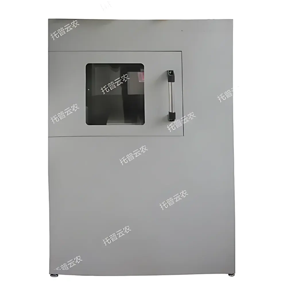
Top Cloud-agri HY-1080 Agricultural X-ray Imaging System for Seed Viability Assessment
| Brand | Top Cloud-agri |
|---|---|
| Origin | Zhejiang, China |
| Manufacturer Type | Direct Producer |
| Product Category | Domestic |
| Model | HY-1080 |
| Instrument Type | X-ray Radiographic Imaging System for Seed Internal Structure Analysis |
| Imaging Sensor Resolution | >1.5 Megapixels |
| Field of View | Ø80 mm |
| Spatial Resolution | 56 lp/cm |
| Output Screen Luminance | >7 cd/cm² |
| Maximum Sample-to-Detector Distance | 300 mm |
| Tube Voltage Range | 60–85 kV |
| Tube Current Range | 0.2–0.4 mA |
| Contrast Sensitivity | 7% |
| Gray Scale Levels | 7-bit |
| Leakage Radiation | <5 mR/h |
| Net Weight | 6.5 kg |
Overview
The Top Cloud-agri HY-1080 Agricultural X-ray Imaging System is a dedicated non-destructive radiographic instrument engineered for high-contrast internal structural analysis of intact seeds. Based on low-dose transmission X-ray imaging principles, it enables direct visualization of embryonic morphology, endosperm integrity, cavity formation (e.g., insect boreholes or developmental voids), and tissue density gradients—parameters strongly correlated with physiological seed viability, germination potential, and developmental maturity. Unlike conventional germination assays requiring days to weeks, this system delivers objective, quantitative morphological data within seconds per sample. Its compact, self-shielded architecture complies with IEC 61331-1 for portable X-ray equipment, incorporating lead-equivalent shielding and interlocked exposure control to ensure operator safety during field-deployable or laboratory-based operation.
Key Features
- Real-time digital radiography without darkroom requirements: Integrated high-luminance output screen (>7 cd/cm²) and >1.5-megapixel CMOS imaging sensor enable immediate visual assessment and frame capture.
- USB 2.0 interface for lossless digital image export: Supports DICOM-compliant TIFF/PNG output for archival, comparative analysis, and integration into seed quality management databases.
- Adjustable imaging parameters: Tube voltage (60–85 kV) and current (0.2–0.4 mA) are optimized per seed species and size (e.g., cereals vs. tree nuts); software-based contrast enhancement and gray-level mapping (7-level scale) improve differentiation of subtle density variations.
- Portable, ruggedized design: Total mass of 6.5 kg with integrated carrying case, foldable collimator, and shock-absorbing chassis—validated for transport between seed testing labs, quarantine stations, and field collection sites.
- Comprehensive radiation safety: Meets national occupational exposure limits (<5 mR/h at 5 cm from housing); automatic beam cutoff upon lid opening and redundant timer-based exposure termination.
Sample Compatibility & Compliance
The HY-1080 accommodates a broad spectrum of agricultural and forestry seeds—from small-grain cereals (e.g., rice, wheat) to large dicotyledonous seeds (e.g., soybean, castor, walnut)—within its Ø80 mm field of view. Its spatial resolution (56 line pairs/cm) resolves sub-millimeter embryonic structures, including cotyledon separation, radicle primordia, and vascular bundle continuity. The system supports standardized seed evaluation protocols aligned with ISTA Rules (International Seed Testing Association), particularly Chapter 5 (Radiography) and Annex B (X-ray Image Interpretation Guidelines). All image metadata—including kV/mA settings, exposure time, date/time stamp, and operator ID—are embedded in EXIF headers, satisfying traceability requirements under ISO/IEC 17025-accredited seed testing laboratories.
Software & Data Management
Bundled proprietary acquisition software provides real-time preview, single-frame capture, batch image sequencing, and annotation tools (ROI measurement, magnification, pseudo-color overlay). Images are stored with embedded calibration references and support batch export to CSV for statistical correlation with germination trials (e.g., % viable embryos vs. lab germination rate). Audit trails record all user actions (image deletion, parameter modification, export events), fulfilling documentation rigor expected in GLP-compliant seed certification workflows. Export formats include TIFF (uncompressed), PNG (lossless), and JPEG2000 (wavelet-compressed), compatible with third-party image analysis platforms such as ImageJ/Fiji or MATLAB-based seed phenotyping pipelines.
Applications
- Pre-germination viability screening for breeding programs and germplasm banks.
- Quarantine inspection of imported seeds for concealed pest infestation (e.g., Callosobruchus, Sitophilus) and mechanical damage.
- Quality control in certified seed production—identifying hollow, shriveled, or malformed embryos prior to packaging.
- Research on seed dormancy mechanisms, embryo development kinetics, and stress-induced morphological anomalies (e.g., heat/drought damage).
- Forestry seed lot assessment for reforestation projects where germination testing is impractical due to long dormancy periods.
FAQ
Is the HY-1080 compliant with international radiation safety standards?
Yes—it conforms to IEC 61331-1 (Medical electrical equipment – X-ray equipment for diagnostic radiology) and GBZ 130-2020 (Chinese regulation for X-ray equipment used in non-medical applications), with verified leakage radiation below 5 mR/h.
Can images be integrated into a LIMS or seed quality management system?
Yes—via standard USB mass storage mode or optional SDK for automated API-driven ingestion; metadata fields align with ISTA XML schema for interoperability.
What is the minimum detectable feature size in a seed embryo?
At optimal kV/mA settings and 1:1 geometric magnification, resolvable features are ≥180 µm (based on 56 lp/cm Nyquist limit), sufficient to distinguish coleoptile initiation and scutellum margins in cereal embryos.
Does the system require external calibration phantoms or annual service certification?
No routine phantom calibration is required; built-in reference markers and factory-installed tube stability monitoring ensure consistent contrast response over 5,000+ exposures. Annual verification by an accredited radiation safety officer is recommended per local regulatory practice.
Is training provided for image interpretation according to ISTA guidelines?
Yes—Top Cloud-agri offers on-site or remote workshops covering radiographic terminology, embryo scoring criteria (e.g., “fully developed” vs. “underdeveloped”), and common artifact recognition (e.g., motion blur, edge enhancement artifacts).