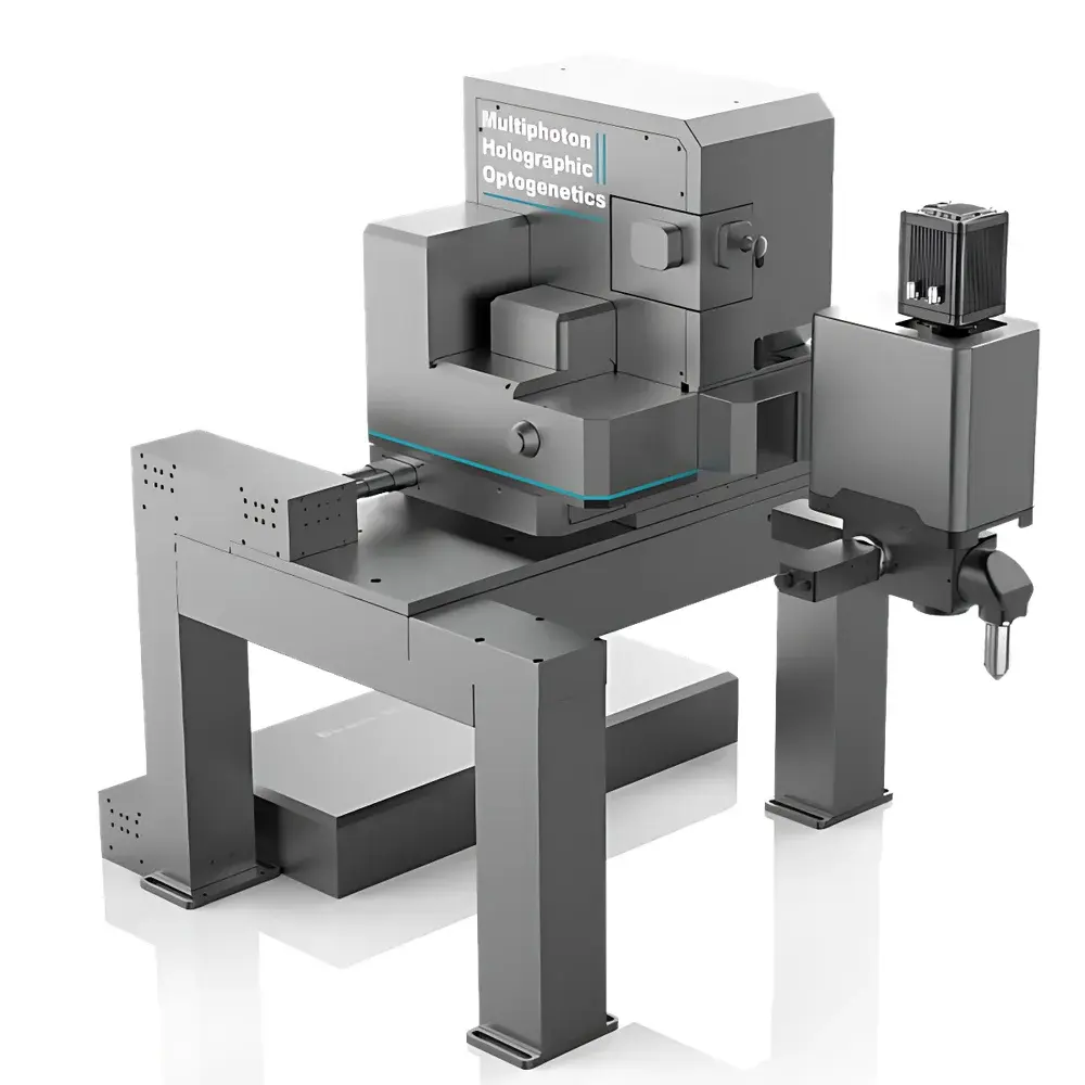
DeepVision Multiphoton Imaging and Holographic Photostimulation System
| Brand | SUSS |
|---|---|
| Origin | Shanghai, China |
| Manufacturer Type | Authorized Distributor |
| Country of Origin | China |
| Model | DeepVision |
| Pricing | Upon Request |
Overview
The DeepVision Multiphoton Imaging and Holographic Photostimulation System is a high-precision, research-grade optical platform engineered for in vivo functional neuroimaging and targeted optogenetic perturbation in intact mammalian tissue. Built upon established principles of nonlinear optical microscopy—including two-photon excitation (2PEF), three-photon excitation (3PEF), second-harmonic generation (SHG), and third-harmonic generation (THG)—the system enables label-free structural imaging and deep-tissue calcium dynamics recording with cellular resolution. Its optical architecture supports simultaneous multimodal acquisition and volumetric photostimulation across millimeter-scale tissue volumes, making it particularly suited for longitudinal studies of neural circuit dynamics, tumor-immune interactions, and pharmacokinetic profiling in live animal models.
Key Features
- Large-sample ergonomic design:龙门-style gantry frame accommodates non-human primates (e.g., macaques) and integrates seamlessly with behavioral apparatuses such as spherical treadmill systems for head-fixed locomotion experiments.
- Extended penetration depth: Three-photon imaging achieves effective signal detection beyond 1500 µm in scattering brain tissue—sufficient to resolve spontaneous and evoked calcium transients from hippocampal CA1 pyramidal neurons in awake, head-restrained mice.
- Multi-regional synchronous imaging & stimulation: Single-field-of-view coverage enables concurrent functional imaging or holographic photostimulation across anatomically distinct brain regions (e.g., prefrontal cortex, thalamus, and hippocampus), supporting causal interrogation of inter-regional connectivity.
- Volumetric holographic optogenetics: Utilizes spatial light modulation (SLM)-based wavefront shaping to generate diffraction-limited excitation foci at user-defined 3D coordinates—enabling independent, subcellular-resolution photostimulation of ≥100 neurons within a single imaging volume.
- Label-free harmonic contrast imaging: SHG and THG modalities provide intrinsic contrast from endogenous structures—including collagen fibrils, myelin sheaths, lipid membranes, and cell boundaries—without exogenous fluorophore labeling or phototoxicity concerns.
Sample Compatibility & Compliance
The DeepVision system is validated for use with diverse biological specimens including murine and non-human primate brains, zebrafish larvae, Drosophila pupae, C. elegans, organoids (cerebral, intestinal, tumor-derived), and vascularized tissue explants. All optical paths comply with IEC 60825-1:2014 Class 4 laser safety standards; mechanical stages meet ISO 10360-2 geometric accuracy specifications. The system supports GLP-compliant experimental workflows through hardware-synchronized timestamping, metadata-embedded image acquisition, and audit-trail-ready configuration logging—facilitating alignment with FDA 21 CFR Part 11 and ISO/IEC 17025 documentation requirements for regulated preclinical research.
Software & Data Management
Control and analysis are unified under the DeepVision Acquisition Suite—a modular, Python-based software environment supporting real-time acquisition, adaptive scanning, and closed-loop feedback control. Image data are stored in standardized OME-TIFF format with embedded OMERO-compatible metadata (including laser power, dwell time, z-step, channel calibration). The suite includes native support for HDF5-based large-volume storage, GPU-accelerated motion correction (using iterative cross-correlation), and export pipelines compliant with BIDS and NWB 2.0 neurodata standards. Optional integration with MATLAB and Python (via PyNWB, CaImAn, Suite2p) enables advanced calcium transient deconvolution, spike inference, and connectomic graph analysis.
Applications
- In vivo deep-brain calcium imaging in awake, behaving mice and macaques—including hippocampal CA1, dentate gyrus, and subcortical nuclei.
- Three-color volumetric imaging of microglia (GFP), vasculature (tdTomato), and neuronal somata (jRGECO1a) in cortical layers L2–L6.
- Label-free THG imaging of brain organoid cytoarchitecture and developmental lamination—without fixation or staining.
- Simultaneous multi-region photoinhibition and photostimulation during associative learning tasks in VR-enabled paradigms.
- Longitudinal tracking of immune cell infiltration and vascular remodeling in orthotopic glioblastoma models using SHG/THG co-registration.
FAQ
What laser sources are integrated into the DeepVision system?
The system is configured with tunable femtosecond lasers (680–1300 nm) optimized for 2PEF, 3PEF, SHG, and THG—each independently adjustable in wavelength, pulse width, and average power.
Is the system compatible with existing optogenetic actuators (e.g., ChRmine, stGtACR2)?
Yes—spectral output and pulse energy profiles are calibrated to match the action spectra and kinetics of modern ultra-sensitive opsins, including red-shifted and bi-stable variants.
Can DeepVision be integrated into an existing electrophysiology or behavioral rig?
The system provides TTL, analog, and Ethernet interfaces for hardware synchronization with patch-clamp amplifiers, DAQ systems, and VR controllers—enabling precise temporal alignment of imaging, stimulation, and behavioral event markers.
Does the software support automated ROI selection and calcium trace extraction?
Yes—the Acquisition Suite includes semi-automated segmentation tools based on constrained non-negative matrix factorization (CNMF-E), with optional cloud-based batch processing via secure API endpoints.
What maintenance and service options are available internationally?
SUSS offers global service contracts including remote diagnostics, annual optical alignment verification, and on-site preventive maintenance—performed by certified field application engineers trained at the Shanghai R&D center.


