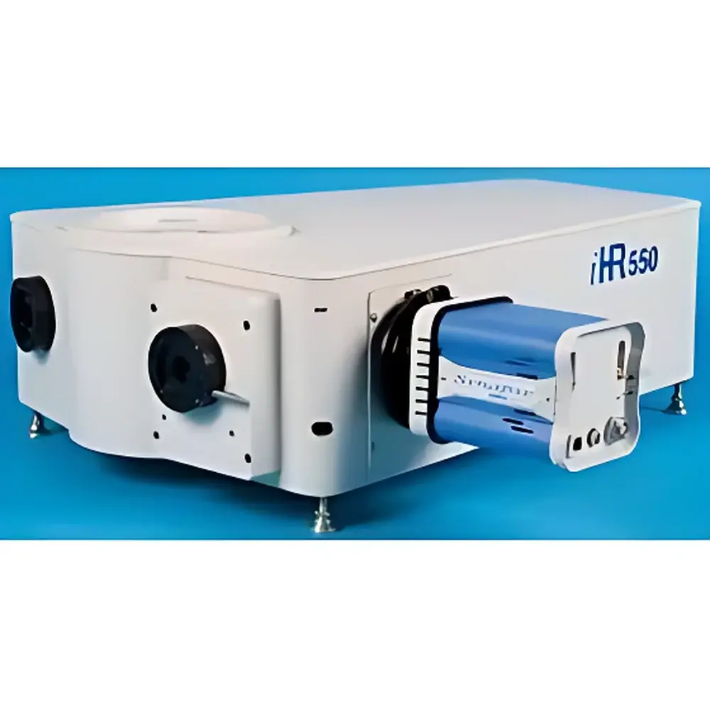
HORIBA Jobin Yvon iHR320 and iHR550 Imaging Spectrographs
| Brand | HORIBA |
|---|---|
| Origin | France |
| Model | iHR320 / iHR550 |
| Application System | Fluorescence Spectroscopy |
| Grating Mount | Dual-grating turret |
| Stray Light | 1.5×10⁻⁴ (iHR320) / 1×10⁻⁵ (iHR550) |
| Focal Length | 320 mm (iHR320), 550 mm (iHR550) |
| f-number | f/4.1 (iHR320), f/6.4 (iHR550) |
| Spectral Resolution | 0.06 nm (iHR320), 0.025 nm (iHR550) |
| Linear Dispersion | 2.31 nm/mm (iHR320), 1.34 nm/mm (iHR550) |
| Grating Size | 68 mm × 68 mm (iHR320), 76 mm × 76 mm (iHR550) |
Overview
The HORIBA Jobin Yvon iHR320 and iHR550 are high-performance, Czerny–Turner imaging spectrographs engineered for demanding fluorescence, Raman, photoluminescence, and time-resolved spectroscopy applications. Designed with precision optical architecture and optimized mechanical stability, these instruments deliver diffraction-limited imaging across the full focal plane—enabling quantitative spectral mapping without vignetting or spatial distortion. Unlike conventional monochromators, the iHR series employs an asymmetric optical layout combined with on-axis grating scanning to maintain constant beam geometry throughout wavelength tuning. This architecture minimizes coma, astigmatism, and higher-order aberrations while preserving throughput and spectral fidelity. The spectrographs utilize toroidal mirrors for astigmatism correction and feature oversized focusing optics to ensure uniform illumination across the entire detector active area—critical for CCD/EMCCD-based hyperspectral imaging and multi-channel detection.
Key Features
- Aberration-Corrected Imaging Optics: Toroidal collimating and focusing mirrors eliminate astigmatism; asymmetric Czerny–Turner configuration reduces coma and field curvature across the flat-field focal plane.
- On-Axis Grating Scanning: Patented grating rotation mechanism maintains incident light at the grating center during wavelength scanning—maximizing diffraction efficiency, minimizing polarization dependence, and ensuring long-term spectral reproducibility.
- Stray Light Suppression: Optimized baffling, blackened internal surfaces, and ray-traced optical path design achieve stray light levels of ≤1.5×10⁻⁴ (iHR320) and ≤1×10⁻⁵ (iHR550), essential for low-signal fluorescence and Raman measurements.
- Modular Flexibility: Dual-input/dual-output port configurations support fiber-coupled, free-space, or microscope-integrated setups; motorized dual-grating turret enables rapid switching between UV-VIS-NIR gratings (e.g., 150–3600 grooves/mm).
- Mechanical Rigidity & Thermal Stability: Monolithic aluminum alloy baseplate with stress-relieved optical mounts; all drive mechanisms undergo metrological validation for repeatability <±0.005 nm over 10⁴ scans.
- CCD-Optimized Interface: Designed in close collaboration with leading detector manufacturers to match pixel pitch, active area, and readout modes—fully compatible with front-illuminated, back-illuminated, deep-depletion, and TE-cooled or LN₂-cooled CCD/EMCCD sensors.
Sample Compatibility & Compliance
The iHR320/iHR550 spectrographs integrate seamlessly into regulated analytical workflows. Their mechanical and software architecture supports audit-trail-enabled operation under GLP and GMP environments when paired with HORIBA’s Synapse or LabSpec 6 software platforms compliant with FDA 21 CFR Part 11 requirements (electronic signatures, user access control, change history logging). Optical performance adheres to ISO 17025 traceable calibration protocols, and spectral accuracy is validated against NIST-traceable emission standards (e.g., Hg/Ar lamps). The instruments accommodate standard 25.4 mm and 50.8 mm optical mounts, vacuum-compatible flanges (optional), and cryogenic sample stages—enabling compatibility with confocal microscopes, low-temperature cryostats, and ultrafast laser excitation systems.
Software & Data Management
Control and data acquisition are managed via HORIBA’s LabSpec 6 software suite, which provides real-time spectral preview, multi-channel synchronization (for gated ICCD or sCMOS detectors), and batch-processing pipelines for spectral deconvolution, peak fitting (Gaussian/Lorentzian/Voigt), and spatial-spectral correlation analysis. Raw data is stored in HDF5 format with embedded metadata (wavelength calibration, grating ID, slit width, integration time, detector temperature), ensuring FAIR (Findable, Accessible, Interoperable, Reusable) compliance. API support (COM, .NET, Python SDK) enables integration into custom automation frameworks—including LabVIEW, MATLAB, and Python-based quantum optics or materials characterization platforms.
Applications
- Microscale fluorescence lifetime imaging (FLIM) and spectral unmixing in biological tissue sections
- Low-frequency Raman mapping of 2D materials (graphene, TMDCs) and strain distribution analysis
- Time-resolved photoluminescence (TRPL) of perovskite thin films and quantum dots
- High-resolution emission spectroscopy of rare-earth-doped phosphors and laser gain media
- Hyperspectral imaging in semiconductor wafer inspection and defect localization
- Coupling with synchrotron beamlines for resonant soft X-ray emission spectroscopy (sXES) post-monochromation
FAQ
What distinguishes the iHR series from conventional monochromators?
The iHR is an imaging spectrograph—not a scanning monochromator—designed to project a spatially resolved spectrum onto a 2D detector. Its flat-field-corrected optics enable simultaneous multi-wavelength detection with minimal spatial distortion.
Can the iHR320/iHR550 be operated under vacuum or purged with inert gas?
Yes—optional vacuum-compatible versions (with fused silica windows and stainless-steel housing) are available for UV applications below 190 nm or for operation in nitrogen-purged or argon-filled environments.
Is grating alignment required after installation or maintenance?
No—the iHR’s kinematic grating mount and factory-aligned optical train eliminate routine realignment; periodic verification using built-in alignment lasers or spectral reference lines is sufficient for ISO 17025-compliant labs.
How does on-axis grating scanning improve measurement reproducibility?
By maintaining constant beam incidence angle and position on the grating surface, on-axis scanning eliminates wavelength-dependent efficiency drift and polarization artifacts—key for quantitative intensity calibration across broad spectral ranges.
Which detector interfaces are supported out-of-the-box?
Standard support includes USB 3.0, Camera Link, and PCIe interfaces for HORIBA Synapse CCDs, Andor iKon-L, Princeton Instruments PI-MAX4 ICCDs, and Hamamatsu ORCA-Fusion BT sCMOS cameras—with full trigger synchronization and hardware binning control.
")