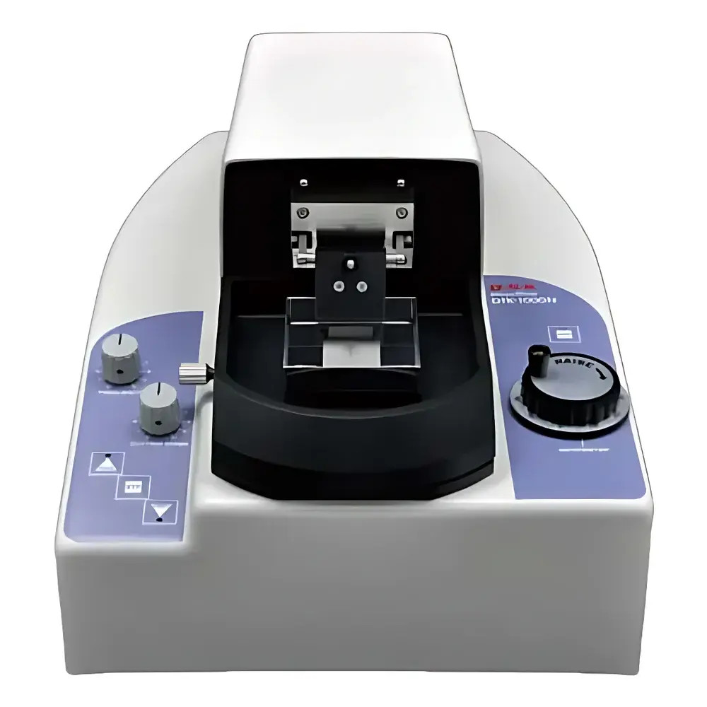
DOSAKA DTK-1000N Vibrating Microtome
| Brand | DOSAKA |
|---|---|
| Origin | Japan |
| Model | DTK-1000N |
| Maximum Sample Dimensions | 25 × 30 mm |
| Slice Thickness Range | 10–500 µm |
| Step Resolution | 1 µm |
| Horizontal Travel | 2 mm |
| Blade Oscillation Frequency | 0–55 Hz |
| Blade Oscillation Amplitude | 2 mm |
| Sectioning Speed | 0–68 mm/min |
| Retraction Speed | 104 mm/min |
| Max Sample Height | 20 mm |
| Compatible Blade Size | 30 mm |
| Power Supply | 220 V, 50 Hz |
| Net Weight | 28 kg |
Overview
The DOSAKA DTK-1000N Vibrating Microtome is an electromechanically driven precision sectioning instrument engineered for the preparation of thick, viable tissue sections from unfixed, non-frozen biological specimens—including freshly excised or chemically fixed neural, cardiac, hepatic, and plant tissues. Unlike cryostat or rotary microtomes, the DTK-1000N employs high-frequency lateral blade oscillation (0–55 Hz) combined with controlled forward advancement to achieve clean, low-compression cuts in soft, hydrated, or elastic samples that would otherwise deform or tear under static cutting forces. This principle—based on resonant harmonic vibration coupled with micrometer-precise specimen advancement—enables reproducible sectioning of fragile brain slices (e.g., hippocampal or cortical preparations) at thicknesses ranging from 10 µm (for electrophysiological recordings) to 500 µm (for organotypic culture or metabolic assays), without requiring cryoprotection, embedding, or dehydration.
Key Features
- Micrometer-adjustable slice thickness control with 1 µm step resolution across a broad operational range (10–500 µm), ensuring compatibility with both light microscopy and functional tissue assays.
- Programmable oscillation frequency (0–55 Hz) and amplitude (2 mm fixed), optimized to match tissue viscoelasticity—reducing chatter artifacts and preserving ultrastructural integrity.
- Motorized horizontal travel (2 mm stroke) with independent retraction speed (104 mm/min), enabling rapid sample repositioning between serial sections while minimizing mechanical drift.
- Stainless-steel 30 mm blade holder with precise angular alignment mechanism, supporting standard disposable or reusable tungsten-carbide blades for consistent edge geometry and longevity.
- Integrated vibration-damping baseplate and rigid aluminum alloy frame, engineered to suppress harmonic coupling during high-frequency operation and maintain positional stability over extended run times.
- Compliance with IEC 61000-6-2 (immunity) and IEC 61000-6-4 (emission) standards for laboratory electromagnetic environments.
Sample Compatibility & Compliance
The DTK-1000N accommodates specimens up to 25 mm (W) × 30 mm (D) × 20 mm (H), making it suitable for rodent brain hemispheres, primate cortical blocks, or intact seedling stems. It supports agarose-embedded, paraformaldehyde-fixed, and fresh vibratome-grade tissue without pre-freezing—preserving endogenous enzyme activity, receptor conformation, and synaptic connectivity. The system meets requirements for GLP-compliant histology workflows when paired with traceable calibration logs and operator-defined SOPs. While not certified as medical device (MDR/IVDR), its mechanical design and material biocompatibility align with ISO 13485-aligned manufacturing practices observed in Class I laboratory instruments.
Software & Data Management
The DTK-1000N operates via front-panel membrane keypad with real-time LED feedback; no proprietary software is required for basic operation. However, optional RS-232 or USB-to-serial interface enables integration into centralized lab automation platforms for audit-trail logging of sectioning parameters (frequency, thickness, speed, timestamp). When used in regulated environments (e.g., preclinical neuropharmacology labs), parameter change history can be exported for inclusion in 21 CFR Part 11–compliant electronic records, provided external validation protocols are implemented per institutional QA policy.
Applications
- Preparation of live brain slices for patch-clamp electrophysiology and calcium imaging.
- Sectioning of fixed spinal cord or retinal tissue for immunohistochemical profiling with antigen preservation.
- Generation of thick sections (100–300 µm) for ex vivo metabolic flux studies using Seahorse or similar platforms.
- Plant tissue sectioning for vascular bundle analysis without artifact-inducing fixation.
- Serial section reconstruction for 3D electron microscopy sample preparation (when combined with post-section staining and resin infiltration).
FAQ
Is the DTK-1000N compatible with cryoprotected or frozen tissue?
No—it is specifically designed for unfrozen, hydrated specimens. Frozen or OCT-embedded tissue requires a cryostat microtome.
What blade types are recommended for optimal performance?
Standard 30 mm stainless-steel or tungsten-carbide blades (e.g., Ted Pella #11000 or Electron Microscopy Sciences #72000) are validated for use; diamond knives are not supported.
Can slice thickness be changed mid-run?
Yes—the micrometer adjustment allows real-time modification without interrupting the sectioning cycle, provided the specimen remains securely clamped.
Does the unit include a cooling stage or perfusion chamber?
No—these are optional accessories; the base model operates at ambient temperature and requires external chilled saline or ACSF delivery systems for physiological viability.
Is technical support available outside Japan?
Yes—authorized service partners provide calibration, preventive maintenance, and on-site repair in North America, EMEA, and APAC regions under DOSAKA’s global distributor network.


