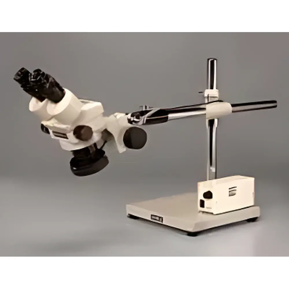
RWD DV-6500 Series Dual- or Trinocular Zoom Stereo Microscope
| Brand | RWD |
|---|---|
| Origin | Japan |
| Manufacturer Type | OEM Manufacturer |
| Product Origin | Imported |
| Model | DV-6500 Series |
| Price | Upon Request |
| Zoom Ratio | 6.5:1 |
| Zoom Range | 0.7× – 4.5× |
| Total Magnification (Standard) | 7× – 45× (with 10× eyepieces, no auxiliary lens) |
| Field of View | 32 mm – 5.1 mm |
| Working Distance (Standard) | 93 mm (10× eyepieces, no auxiliary lens) |
| Binocular Tube Inclination | 60° |
| Trinocular Tube Inclination | 45° |
| Interpupillary Adjustment Range | 54 mm – 75 mm |
| Diopter Adjustment Range | ±5 D per eyepiece tube |
| Extended Magnification Range (Dual) | 2.1× – 270× (with optional 2.0× auxiliary lens and 30× eyepieces) |
| Extended Magnification Range (Trino) | 0.98× – 203× (with optional 1.5× auxiliary lens and 30× eyepieces) |
| Extended Working Distance Range (Dual) | 34 mm – 251 mm (with 2.0× to 0.3× auxiliary lenses) |
| Extended Working Distance Range (Trino) | 38 mm – 300 mm (with 0.28× to 1.5× auxiliary lenses) |
| Eyepiece Options | 5×, 10× (graduated), 15×, 20×, 30× |
| Auxiliary Lens Options | 0.28×, 0.3×, 0.35×, 0.44×, 0.5×, 0.75×, 1.0×, 1.5×, 2.0× |
| Illumination Compatibility | LED ring light, cold light source, fluorescence excitation modules |
| Imaging Interface | C-mount adapter (trinocular), USB 2.0 digital camera included in trinocular configuration |
| Mounting Flexibility | Universal articulated stand (610 mm height), sliding lens carrier, 360° rotating head |
Overview
The RWD DV-6500 Series Dual- or Trinocular Zoom Stereo Microscope is an optomechanically engineered instrument designed for high-fidelity stereoscopic observation across life science, preclinical research, and precision industrial inspection applications. Based on the Greenough optical design principle—featuring two separate optical paths with angled objectives—the system delivers true depth perception, minimal parallax, and high-resolution three-dimensional visualization. Its continuously variable zoom optics (6.5:1 ratio) enable seamless magnification transitions from low-power macroscopic survey (0.7×) to detailed microstructural analysis (4.5×), while maintaining focus stability and image flatness across the entire field. The microscope’s modular architecture supports both dual-eyepiece (binocular) and trinocular configurations, with a fully rotatable head (360°) and ergonomic tube inclination (60° binocular / 45° trinocular) to accommodate prolonged user sessions under GLP-compliant laboratory conditions.
Key Features
- High-stability Greenough-style stereo optical path with matched objective pairs ensuring consistent resolution and depth of field
- Continuously adjustable zoom mechanism with backlash-free gear train and calibrated magnification scale
- Wide interpupillary adjustment (54–75 mm) and independent ±5 D diopter compensation per eyepiece tube for operator-specific visual acuity correction
- Robust universal mounting platform with 610 mm-height articulated stand, precision sliding lens carrier, and vibration-damped base
- Integrated compatibility with multiple illumination modalities: LED ring illuminator (cool white, uniform intensity), fiber-optic cold light sources, and optional fluorescence excitation modules for live-tissue contrast enhancement
- Trinocular port equipped with standard C-mount interface and factory-calibrated USB 2.0 digital imaging system (2 MP resolution, real-time capture, TIFF/AVI export)
- Modular auxiliary lens system supporting working distances from 34 mm to 300 mm and extended magnifications up to 270× (dual) or 203× (trino)
Sample Compatibility & Compliance
The DV-6500 accommodates diverse specimen types—including live small animals (mouse, rat, zebrafish), dissected tissues, botanical samples, microelectronics assemblies, and metallurgical cross-sections—without requiring vacuum or conductive coating. Its long working distance (standard 93 mm; extendable to 300 mm) permits unobstructed access for micromanipulators, surgical tools, or pipetting systems during in situ procedures. The system complies with ISO 10934-1:2004 (optical microscopy—nomenclature and terminology) and meets mechanical safety requirements per IEC 61010-1:2010 for laboratory equipment. When configured with audit-trail-enabled imaging software (optional), it supports FDA 21 CFR Part 11 compliance for electronic records and signatures in regulated environments. All optical components are anti-reflection coated (MgF₂ multilayer) to maximize light transmission and minimize chromatic aberration across visible wavelengths (400–700 nm).
Software & Data Management
The included RWD ImageCapture Pro software provides full control over exposure time, white balance, gain, and gamma correction via USB 2.0 interface. Captured images and time-lapse sequences are stored with embedded metadata (magnification, date/time stamp, objective/lens configuration, user ID). Export formats include uncompressed TIFF (for quantitative analysis), JPEG (reporting), and AVI (dynamic process documentation). Optional RWD Analysis Suite adds measurement tools (line, angle, area, particle count), annotation layers, and batch-processing workflows compatible with common LIMS and ELN platforms. All software installations generate immutable log files for GLP/GMP traceability and are validated for use in ISO 17025-accredited testing laboratories.
Applications
- Neurosurgical procedures and stereotaxic brain mapping (compatible with standard rodent stereotactic frames)
- Intraoperative guidance during microvascular anastomosis and nerve repair
- Developmental biology studies involving embryo dissection and organ explant culture
- Quality assurance in PCB assembly, solder joint inspection, and semiconductor packaging
- Forensic evidence examination (fiber, hair, toolmark analysis)
- Educational demonstration of anatomical structures and histological tissue relationships
- Materials science evaluation of fracture surfaces, coating uniformity, and grain morphology
FAQ
What is the maximum usable magnification without significant resolution loss?
The practical upper limit for high-fidelity imaging is ~120× when using 30× wide-field eyepieces with a 1.5× auxiliary lens and optimal illumination—beyond this, diffraction limits and depth-of-field constraints reduce interpretive utility.
Can the microscope be integrated into an automated workflow?
Yes—via TTL-compatible shutter triggers and RS-232/USB command protocols, the system supports synchronization with motorized stages, environmental chambers, and third-party acquisition software (e.g., MATLAB, Python OpenCV APIs).
Is calibration certification available for metrological applications?
RWD provides NIST-traceable calibration certificates (per ISO/IEC 17025) for magnification accuracy, field diameter, and working distance upon request—valid for 12 months from issuance.
How is thermal drift managed during extended imaging sessions?
The all-metal optical housing (aluminum alloy chassis with brass focusing gears) features low coefficient of thermal expansion; combined with passive heat-sink design in the LED illuminator, it maintains optical alignment stability within ±0.5 µm over 4-hour continuous operation.
Are replacement parts and service support available globally?
RWD maintains authorized service centers in North America, EU, and APAC regions; all optical components carry a 3-year limited warranty, and firmware updates are distributed quarterly through secure customer portal access.


