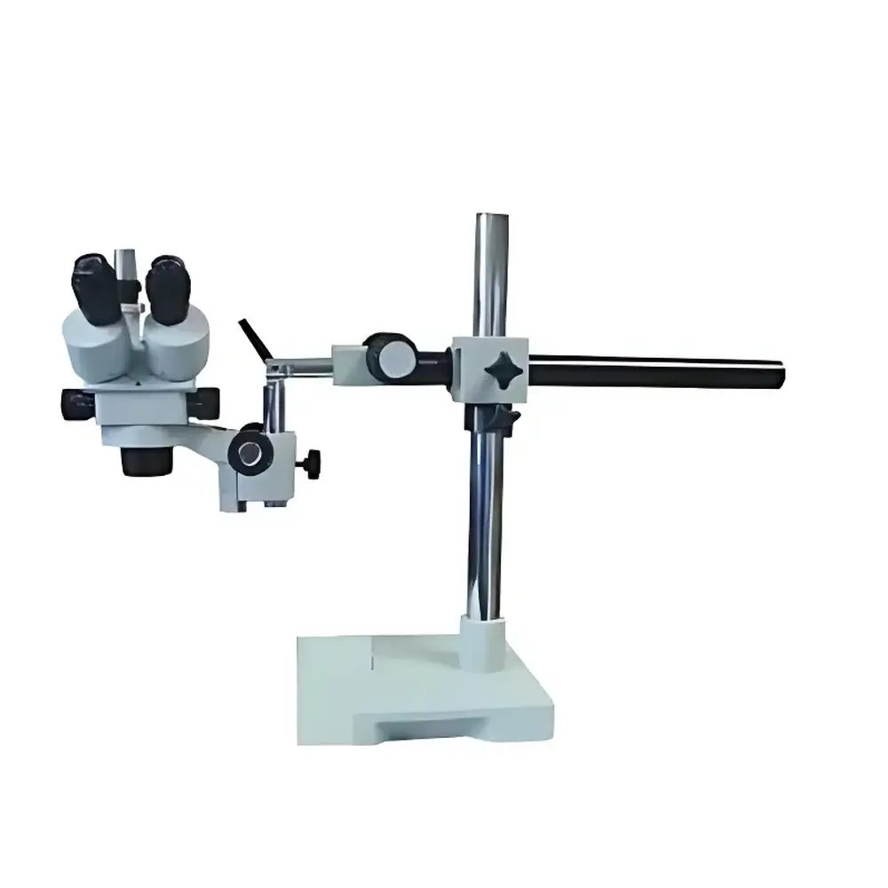
RWD 77001 Desktop Stereo Microscope with 0.5× Auxiliary Lens
| Brand | RWD |
|---|---|
| Origin | Shenzhen, China |
| Manufacturer Type | Direct Manufacturer |
| Origin Category | Domestic (China) |
| Model | 77001 |
| Price | USD 1,120 (approx. based on ¥8,128 at 1 USD ≈ ¥7.26) |
| Eyepiece | Widefield 10× (WF10×) |
| Zoom Objective Range | 0.7–4.5× |
| Zoom Ratio | 6.5:1 |
| Optical Magnification | 7–45× (standard), up to 7–225× with optional accessories |
| Interpupillary Distance Adjustment | 55–75 mm |
| Vertical Travel (Column Height Adjustment) | 450 mm |
| Horizontal/Vertical Stage Travel | 0–500 mm |
| Working Distance | ≥110 mm at lowest magnification (0.7×) |
| Auxiliary Lens | Integrated 0.5× lens for extended field of view and reduced magnification |
| Illumination | Dual-mode (top LED reflected + bottom LED transmitted), adjustable intensity |
Overview
The RWD 77001 Desktop Stereo Microscope is an ergonomically engineered dual-eye stereoscopic imaging system designed for high-fidelity visual inspection, microsurgical preparation, and precision biological dissection. Based on the Greenough optical principle—employing two separate, parallel optical paths—the instrument delivers true binocular depth perception and a naturally oriented, upright 3D image without inversion or mirror reversal. This architecture ensures minimal visual fatigue during prolonged observation sessions and supports accurate spatial interpretation essential for neuroanatomical tracing, embryonic tissue manipulation, small-animal surgical staging, and histological specimen survey. The system integrates a continuously variable zoom objective (0.7×–4.5×) coupled with widefield 10× eyepieces, yielding a standard optical magnification range of 7×–45×. With the included 0.5× auxiliary lens, the effective magnification extends downward to 3.5×, significantly increasing field diameter and working distance—critical for macro-to-micro transitional workflows in preclinical labs.
Key Features
- Continuous zoom optics with 6.5:1 zoom ratio (0.7×–4.5×), enabling seamless magnification transitions without discrete step changes or lens swapping
- Widefield 10× eyepieces (WF10×) providing extended eye relief and uniform edge-to-edge sharpness across the entire field of view
- Adjustable interpupillary distance (55–75 mm) accommodating diverse user anthropometry and ensuring optimal ocular alignment
- Motor-free mechanical stage with 0–500 mm total travel range (X-Y-Z), supporting large specimen handling and multi-position repositioning without refocusing
- 450 mm vertical column lift capacity, facilitating rapid height adjustment for varying sample thicknesses and user posture requirements
- Dual-illumination system: top-mounted coaxial LED for reflected-light surface inspection and bottom-transmitted LED for translucent or semi-opaque specimens—both independently dimmable via rotary controls
- Integrated 0.5× auxiliary lens mounted in fixed optical path, eliminating alignment drift and preserving parfocality during low-magnification panoramic scanning
Sample Compatibility & Compliance
The RWD 77001 accommodates a broad spectrum of biological and material samples—including live rodent tissues, zebrafish embryos, plant meristems, insect dissections, PCB assemblies, and polymerized hydrogels—without requiring vacuum or conductive coating. Its ≥110 mm working distance at minimum magnification permits unobstructed access for micromanipulators, forceps, and pipette positioning. The microscope complies with IEC 61000-6-3 (EMC emission standards) and IEC 61000-6-2 (immunity), and its LED illumination meets IEC 62471 photobiological safety Class 1 requirements. While not certified for ISO 13485 or FDA 21 CFR Part 11 out-of-the-box, its stable optical output, repeatable mechanical positioning, and non-volatile illumination settings support GLP-aligned documentation protocols when integrated into validated laboratory workflows.
Software & Data Management
The RWD 77001 operates as a hardware-optimized optical platform without proprietary firmware or embedded software. It is fully compatible with third-party digital imaging solutions: standard C-mount adapters (0.5× or 1×) enable integration with USB 3.0 CMOS cameras (e.g., Thorlabs DCC1545M, Lumenera Lt545) and machine vision software suites including NIS-Elements (Nikon), ZEN Blue (Zeiss), and open-source platforms such as Micro-Manager 2.0. All image capture, annotation, measurement (distance, area, angle), and time-lapse sequencing are managed externally—ensuring full audit trail traceability, DICOM-compliant metadata embedding, and compatibility with LIMS environments. No cloud connectivity or remote control firmware is present, aligning with air-gapped lab security policies.
Applications
- Preclinical surgical planning and intraoperative guidance in rodent models of stroke, spinal cord injury, and tumor xenograft resection
- Developmental biology: real-time observation of avian/chick embryo vasculogenesis, Xenopus gastrulation, and Drosophila larval CNS patterning
- Botanical morphology: leaf epidermal stomatal analysis, root hair quantification, and fungal hyphal growth tracking
- Quality assurance in microelectronics: solder joint inspection, flex-circuit defect mapping, and MEMS device verification
- Educational laboratories: comparative anatomy instruction, forensic fiber analysis, and materials science demonstration of fracture propagation
FAQ
Is the 0.5× auxiliary lens permanently installed or interchangeable?
The 0.5× auxiliary lens is factory-integrated into the main optical train and cannot be removed without recalibration. It is mechanically locked to preserve parfocality and axial alignment across the full zoom range.
What is the maximum usable field diameter at 7× magnification with the 0.5× lens?
At 7× (0.7× objective × 10× eyepiece × 0.5× auxiliary), the nominal field number is 22 mm, yielding a ~31.4 mm field diameter—verified per ISO 8578:2017 methodology.
Can this microscope be adapted for fluorescence observation?
No. The optical path lacks dichroic beam splitters, excitation/emission filter slots, or UV-transmissive glass elements; it is strictly optimized for brightfield and oblique white-light contrast.
Does RWD provide calibration certificates or metrological traceability documentation?
Calibration reports for magnification accuracy and stage linearity are available upon request (ISO/IEC 17025-accredited third-party verification optional at additional cost).
Is the column height adjustment motorized or manual?
Height adjustment is manually operated via a precision rack-and-pinion mechanism with locking collar—designed for stability under load and resistance to thermal drift.


