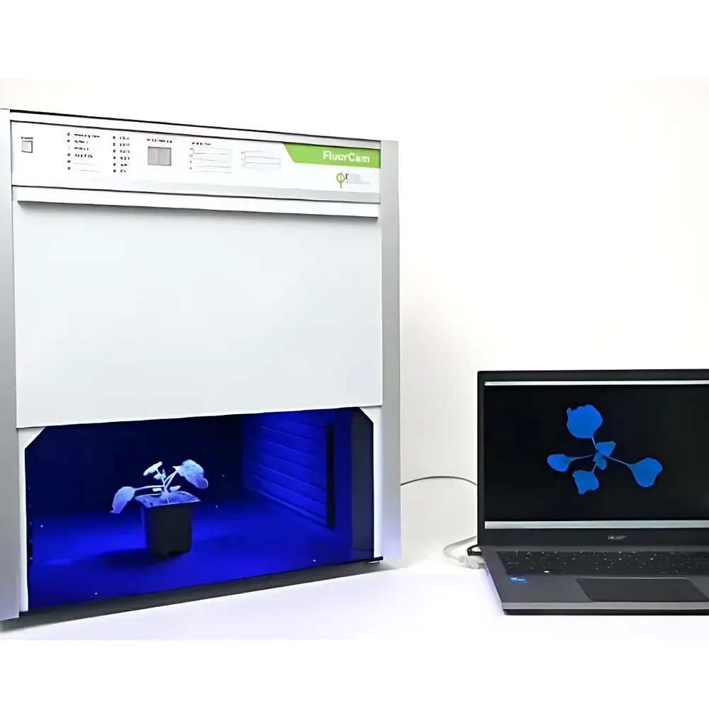
PSI FluorCam Closed-System GFP/Chlorophyll Fluorescence Imaging System
| Brand | PSI (Photosynthesis Systems Instruments) |
|---|---|
| Origin | Czech Republic |
| Model | FluorCam Closed-System |
| Imaging Area | 13 × 13 cm |
| Sensor | TOMI-2 high-resolution CCD (1360 × 1024 px, 16-bit A/D, 20 fps max) |
| Light Sources | 4 LED panels (2 × red-orange, 2 × blue) |
| Filter Wheel | 7-position motorized, software-controlled with Chl *a* and GFP filter sets |
| Fluorescence Parameters | >50 chlorophyll fluorescence parameters (Fo, Fm, Fv/Fm, NPQ, qP, ETR, Y(NO), Y(NPQ), Rfd, etc.), all exportable as spatially resolved false-color images |
| Optional Modules | OJIP fast kinetics imaging, QA⁻ reoxidation kinetics, PAR absorption & NDVI imaging, temperature-controlled sample stage (15–75 °C) |
| Software | FluorCam v8.x with protocol editor, time-stamped automated acquisition, GLP-compliant data logging, and multi-parameter image overlay |
Overview
The PSI FluorCam Closed-System GFP/Chlorophyll Fluorescence Imaging System is a fully integrated, dark-adapted platform engineered for quantitative, spatially resolved dual-channel fluorescence phenotyping of live plant specimens. It combines pulse-amplitude modulation (PAM) chlorophyll *a* fluorescence imaging with high-sensitivity green fluorescent protein (GFP) detection—enabling concurrent assessment of photosynthetic performance and transgene expression in the same biological sample. The system operates on the principle of modulated fluorometry: weak measuring light pulses excite PSII reaction centers while saturating flashes transiently close all reaction centers, allowing precise calculation of quantum yield, electron transport rate (ETR), non-photochemical quenching (NPQ), and other biophysically grounded parameters. GFP detection leverages optimized excitation/emission band separation via motorized 7-position filter wheels and spectrally matched LED sources, ensuring minimal crosstalk between chlorophyll and GFP channels. Designed for reproducible, operator-independent measurements, the closed-system architecture eliminates ambient light interference and standardizes dark adaptation—critical for reliable Fo and Fv/Fm determination per ISO 10211 and ASTM E2917 protocols.
Key Features
- Fully enclosed dark-adaptation chamber with integrated LED illumination, eliminating external light contamination and enabling standardized pre-measurement acclimation.
- Simultaneous dual-channel acquisition: PAM-based chlorophyll *a* fluorescence and GFP fluorescence within a single imaging session—no manual repositioning or hardware switching required.
- High-fidelity TOMI-2 CCD sensor: 1360 × 1024 resolution, 16-bit dynamic range (65,536 gray levels), and 20 fps video capture at full resolution for kinetic analysis of rapid fluorescence transients (e.g., OJIP, QA⁻ reoxidation).
- Configurable LED light engine: Four independently controlled high-power LED panels (standard: 2 red-orange + 2 blue); actinic irradiance scalable to 3000 µmol·m⁻²·s⁻¹; saturation flash up to 6000 µmol·m⁻²·s⁻¹.
- Motorized 7-position filter wheel with factory-calibrated Chl *a* and GFP filter sets; additional positions support user-defined fluorophores (YFP, BFP, RFP, DAPI) under software control.
- Comprehensive protocol library: Editable experimental workflows for Fv/Fm, Kautsky induction, light-response curves (LC), quenching analysis, NDVI/PAR absorption, and time-series monitoring—with automated timestamped data archiving.
- Optional advanced modules: OJIP fast fluorescence kinetics (20+ derived parameters), QA⁻ reoxidation dynamics, and programmable temperature control (15–75 °C, ±0.2 °C stability) via 48-well heated stage.
Sample Compatibility & Compliance
The FluorCam Closed-System accommodates diverse biological formats without modification: detached leaves, intact seedlings, whole rosettes, mosses, lichens, algal suspensions, and microtiter plates (96-/384-well). Its 13 × 13 cm field-of-view supports both high-throughput screening and detailed organ-level mapping. All fluorescence parameters comply with internationally recognized definitions from the International Society of Photosynthesis Research (ISPR) and are traceable to primary literature standards (e.g., Schreiber et al., 1986; Klughammer & Schreiber, 2008). Data output adheres to FAIR principles (Findable, Accessible, Interoperable, Reusable) and supports 21 CFR Part 11-compliant audit trails when deployed in regulated environments (e.g., GLP/GMP-compliant phenotyping labs). Calibration routines include built-in dark-current correction, flat-field normalization, and reference standard validation using certified fluorescence plates.
Software & Data Management
FluorCam v8.x software provides an integrated environment for acquisition, preprocessing, quantification, and visualization. The Protocol Editor enables drag-and-drop construction of multistep experiments—including sequential light treatments, dark intervals, and multi-wavelength acquisitions—with conditional branching and error handling. Raw TIFF stacks are automatically annotated with metadata (timestamp, protocol ID, instrument configuration, environmental logs) and stored in hierarchical folder structures. Batch processing tools support region-of-interest (ROI) extraction, parameter mapping, statistical overlay, and export to CSV, HDF5, or MATLAB-compatible formats. Time-series datasets generate animated GIFs or AVI files for kinetic visualization. For regulatory use, optional software modules provide electronic signatures, user access controls, and immutable audit logs meeting FDA and EMA requirements for analytical instrument qualification.
Applications
- Functional genomics: Spatial correlation of GFP-tagged protein localization with photosynthetic efficiency across mutant libraries or CRISPR-edited lines.
- Abiotic stress phenotyping: Quantitative mapping of drought-, heat-, or salinity-induced heterogeneity in PSII integrity (Fv/Fm decline) and antioxidant reporter expression (e.g., GFP-HSP70).
- Chemical biology: High-content screening of photosynthesis-modulating compounds using dose-dependent OJIP parameter shifts and concurrent GFP-based toxicity reporters.
- Plant-microbe interactions: Dual-channel imaging of pathogen-induced chlorophyll fluorescence suppression alongside defense-related promoter-driven GFP activation.
- Ecophysiology: Comparative analysis of light-use efficiency (ETR vs. PAR) and photoprotective capacity (NPQ kinetics) across ecotypes under controlled spectral regimes.
- Algal biotechnology: Rapid assessment of lipid accumulation strains via chlorophyll fluorescence decline coupled with Nile Red or BODIPY-GFP co-staining.
FAQ
Can the system quantify absolute GFP concentration?
No—it provides relative fluorescence intensity normalized to background and exposure settings. Absolute quantification requires parallel biochemical assays (e.g., ELISA, Western blot) or calibrated reference standards.
Is remote operation supported?
Yes. The system connects via Gigabit Ethernet and supports secure remote desktop access, protocol triggering via HTTP API, and real-time video streaming for off-site monitoring.
What maintenance is required for long-term calibration stability?
Annual verification using NIST-traceable neutral density filters and fluorescence reference tiles is recommended. LED output drift is tracked automatically via internal photodiode feedback; software compensates for minor variations in real time.
Can I integrate third-party environmental sensors (e.g., CO₂, humidity)?
Yes. The FluorCam software accepts analog/digital inputs via optional DAQ modules, enabling synchronized logging of chamber conditions with fluorescence data.
Does the system support machine learning-based phenotypic classification?
Raw TIFF stacks and parameter maps are compatible with Python (scikit-image, TensorFlow) and R (EBImage, phenofit) pipelines. PSI provides documented HDF5 export templates optimized for feature extraction and model training.


