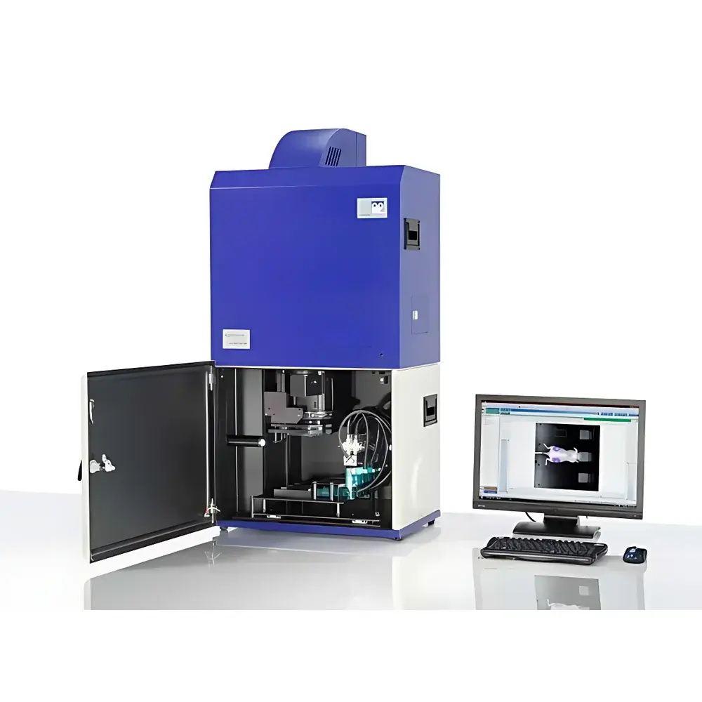
Berthold LB983 NightOWL II Small Animal In Vivo Optical Imaging System
| Brand | Berthold |
|---|---|
| Origin | Germany |
| Model | LB983 |
| Imaging Modality | Bioluminescence & Fluorescence Optical Imaging |
| Camera Type | Cooled Slow-Scan CCD |
| Cooling Method | Peltier (−90 °C typical operating temperature) |
| Spectral Range | 400–900 nm |
| Pixel Resolution | Up to 3.2 MP (NC320 front-illuminated) / 1.0 MP (NC100 back-illuminated) |
| Field of View Adjustment | Motorized vertical camera positioning (35–725 mm travel) |
| Maximum Sample Height | 260 mm |
| Dark Box Light Tightness | < 0.001 lux internal ambient light leakage |
| Calibration | Integrated optical flat-field and intensity reference standard for quantitative cross-experiment comparability |
| PET Integration | MACU interface module with dedicated software synchronization for small-animal PET co-registration |
| Software Platform | IndiGO v5.x with FDA 21 CFR Part 11-compliant audit trail, GLP/GMP-ready reporting modules |
Overview
The Berthold LB983 NightOWL II is a high-sensitivity, cooled slow-scan CCD-based in vivo optical imaging system engineered for quantitative bioluminescence and fluorescence detection in small laboratory animals (mice, rats, zebrafish, and juvenile larger models). Operating on the principle of photon capture from biological light sources—such as luciferase-luciferin reactions (bioluminescence) or exogenous fluorophore excitation (fluorescence)—the system delivers non-invasive, longitudinal molecular readouts without ionizing radiation. Its core architecture integrates a thermoelectrically cooled CCD sensor housed within a light-tight, motorized dark chamber, enabling low-noise, long-exposure acquisition essential for detecting weak signals from deep-tissue reporters. Designed for rigorous preclinical research, the LB983 supports both standalone optical imaging and multimodal workflow integration—specifically via its proprietary MACU (Multi-Modal Acquisition Control Unit) interface—for synchronized data acquisition with small-animal PET scanners. This capability positions the system uniquely within the molecular imaging ecosystem, facilitating correlative analysis across complementary contrast mechanisms while maintaining traceable, reproducible quantification.
Key Features
- Cooled CCD Detection Architecture: Dual-CCD configuration—NC320 front-illuminated (3.2 MP, optimized for fluorescence quantum efficiency up to 85% at 550 nm) and NC100 back-thinned, back-illuminated (1.0 MP, enhanced sensitivity in 500–700 nm range, ideal for low-flux bioluminescence)—both stabilized at ≤ −90 °C via Peltier cooling to suppress dark current and thermal noise.
- Motorized Variable-Focus Dark Chamber: Precision vertical camera translation (35–725 mm range) enables automatic focal plane adjustment for specimens up to 260 mm tall—including whole rodents, tissue explants, microplates, gels, and plant seedlings—ensuring optimal spatial resolution and signal uniformity across heterogeneous sample geometries.
- Quantitative Optical Calibration: Built-in flat-field correction and intensity reference standard ensure pixel-wise radiometric consistency across sessions, instruments, and laboratories—critical for longitudinal studies and multi-center trials requiring inter-experiment comparability.
- Multimodal Integration Ready: MACU hardware interface and synchronized acquisition software allow time-aligned data capture with small-animal PET systems (e.g., Siemens Inveon, Bruker Albira), enabling direct spatial registration of metabolic (PET) and functional/molecular (optical) endpoints without post-hoc image warping.
- Regulatory-Compliant Software Stack: IndiGO v5.x includes full 21 CFR Part 11 compliance features—electronic signatures, role-based access control, immutable audit trails, and version-controlled protocol templates—supporting GLP and GMP-aligned workflows in translational pharmacology and biologics development.
Sample Compatibility & Compliance
The LB983 accommodates diverse biological and material science specimens: live rodents (anesthetized or restrained), zebrafish embryos, cell monolayers, SDS-PAGE/Western/Northern blots, microtiter plates (96-/384-well), thin-layer chromatography strips, polymer films, and botanical tissues. Its optical path supports both epi-illumination (for fluorescence) and trans-illumination (for transmission imaging), with optional filter wheels accommodating standard fluorophores (GFP, RFP, Cy5, IRDye800CW) and chemiluminescent substrates. The system complies with ISO 13485 design controls for medical device-associated research instrumentation and meets electromagnetic compatibility (EMC) requirements per IEC 61326-1. All software modules are validated per ASTM E2500-13 and aligned with USP guidelines for analytical instrument qualification.
Software & Data Management
IndiGO v5.x provides end-to-end image acquisition, processing, and reporting functionality. Key modules include ROI-based radiance quantification (photons/sec/cm²/sr), spectral unmixing for multiplexed fluorophore separation, background subtraction algorithms optimized for low-SNR bioluminescence, and batch-processing pipelines for high-throughput microplate assays. Data export supports DICOM-SR, TIFF (with embedded metadata), CSV, and HDF5 formats. Audit trail logs record every parameter change, user action, and calibration event with timestamp, operator ID, and reason-for-change annotation—fully compliant with FDA and EMA expectations for regulated preclinical data integrity.
Applications
- Longitudinal monitoring of tumor xenograft growth, metastasis, and treatment response using luciferase-expressing cell lines.
- In vivo tracking of stem cell engraftment, migration, and differentiation via lentiviral reporter constructs.
- Pharmacokinetic/pharmacodynamic assessment of fluorescently labeled therapeutics (e.g., antibody-drug conjugates).
- Gene therapy vector biodistribution and promoter activity profiling in transgenic mouse models.
- Neuroinflammation imaging using near-infrared fluorescent probes targeting amyloid-β or activated microglia.
- Real-time visualization of bacterial infection dynamics and antibiotic efficacy in sepsis models.
- High-content screening of reporter gene activity in primary cells or organoids cultured in microplates.
- Chemiluminescent detection of ROS generation in skin disease models or oxidative stress assays.
FAQ
What is the minimum detectable photon flux for bioluminescence imaging?
The NC100 back-illuminated CCD achieves sub-100 photons/sec detection limits under standard 5-minute exposures at −90 °C, depending on lens f-number and emission spectrum.
Can the LB983 perform spectral unmixing with multiple fluorophores simultaneously?
Yes—when equipped with motorized filter wheels and appropriate excitation/emission filters, IndiGO supports linear unmixing of up to four spectrally distinct fluorophores using reference spectra libraries.
Is validation documentation available for GxP environments?
Berthold provides IQ/OQ/PQ protocols, URS templates, and a complete validation support package including risk assessments and test scripts aligned with Annex 11 and ALCOA+ principles.
Does the system support anesthesia integration for live animal imaging?
The LB983 dark chamber includes standardized ports for third-party gas anesthesia delivery systems (e.g., VetEquip, Kent Scientific), with compatible animal holders featuring integrated nose cones and temperature regulation.
How is photobleaching minimized during fluorescence acquisition?
IndiGO implements adaptive exposure control, LED power ramping, and real-time signal monitoring to deliver minimal necessary excitation dose—reducing phototoxicity and preserving sample viability across serial imaging sessions.


