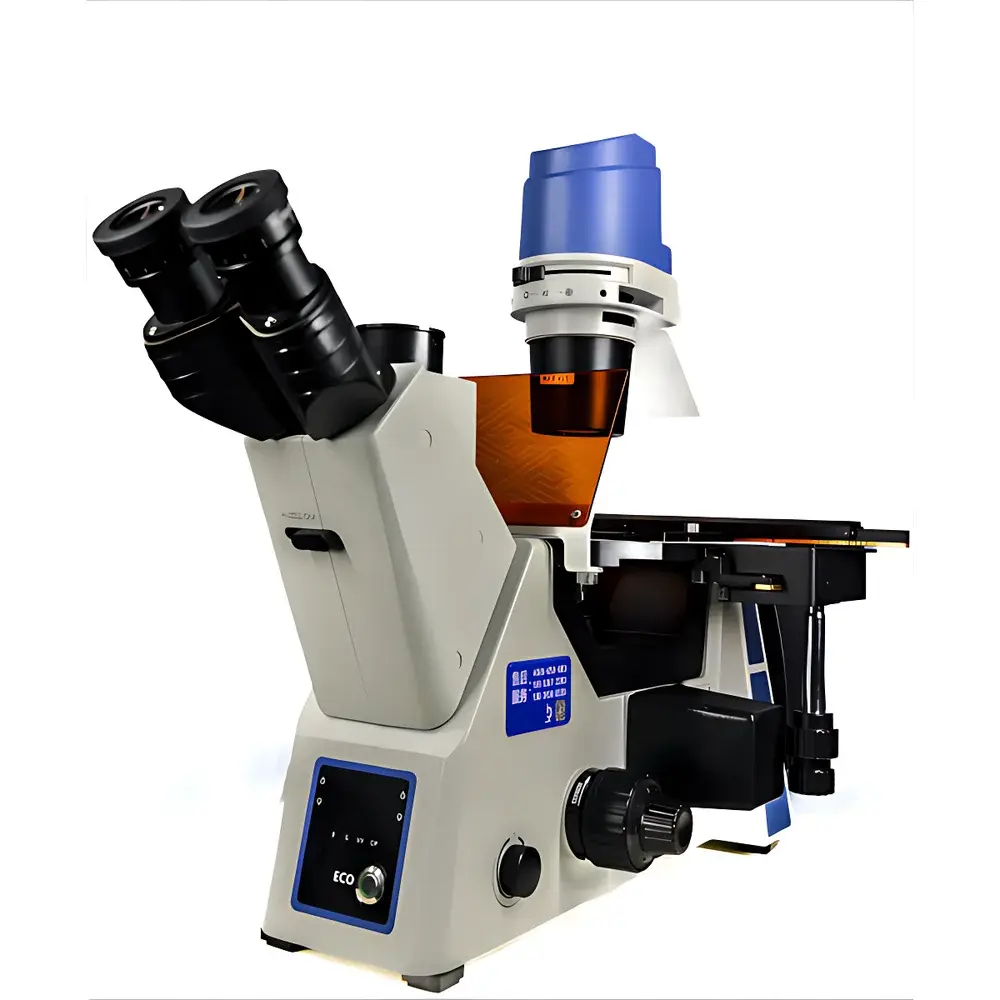
LEI-TECH LK-YG91 Research-Grade Inverted Fluorescence Microscope
| Brand | LEI-TECH |
|---|---|
| Origin | Tianjin, China |
| Manufacturer Type | Original Equipment Manufacturer (OEM) |
| Instrument Category | Inverted Fluorescence Microscope |
| Model | LK-YG91 |
| Excitation Source | High-Stability LED |
| Microscope Class | Research-Grade |
| Eyepieces | Widefield Plan Eyepieces PL10X/22mm |
| Objective Lenses | 4X, 10X, 20X, 40X (Infinity-Corrected) |
| Fluorescence Filter Sets | B1 (Blue Excitation), G1 (Green Excitation) |
| Illumination | Dual-Mode LED (Transmitted & Epi-Fluorescence) |
| Focusing Mechanism | Low-Position Coaxial Coarse/Fine Focus with Anti-Drift Adjustment |
| Condenser | 72 mm Achromatic Condenser with Centering Knobs |
| Observation Head | Trinocular Ergonomic Head, 45° Inclination, 360° Rotatable Hinged Binocular Tube, Interpupillary Distance 50–75 mm, Dual Light Path Splitting (100:0 / 0:100) |
| Stage | Mechanical XY Translation Stage with Extension Plate Option |
| Fluorescence Accessories | Green Contrast Filter, Color Temperature Conversion Filter |
| Camera Interface | C-Mount, Compatible with 20 MP CMOS Sensor (5440 × 3648 px) |
Overview
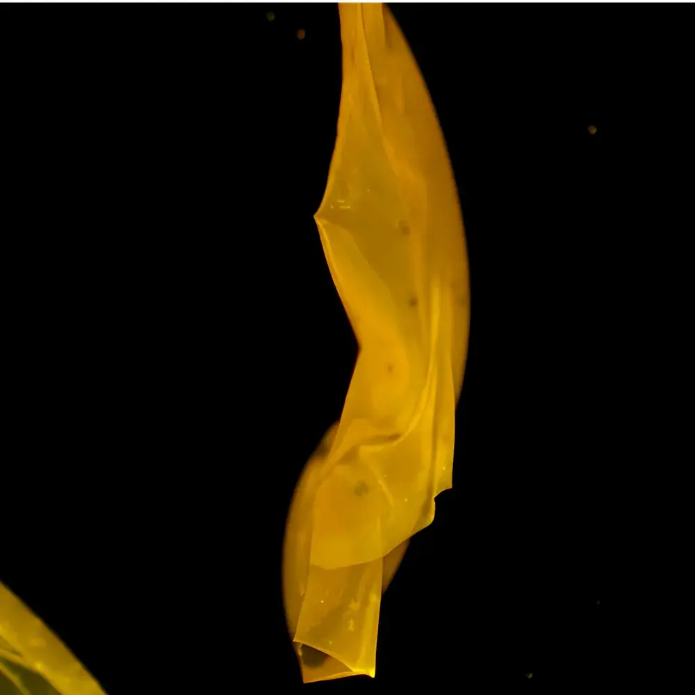
The LEI-TECH LK-YG91 is a research-grade inverted fluorescence microscope engineered for high-fidelity live-cell imaging, long-term tissue culture observation, and multimodal optical analysis in academic laboratories, biotech R&D facilities, and preclinical research centers. Its optical architecture is based on an infinity-corrected dual-compensation optical pathway (UISC), ensuring minimal chromatic and spherical aberration across the full magnification range (40×–400×). Unlike finite-conjugate systems, the UISC design enables stable parfocality, consistent image brightness, and seamless integration of auxiliary modules—including phase contrast sliders, fluorescence filter cubes, and digital imaging interfaces—without compromising resolution or signal-to-noise ratio. The system supports three primary contrast modalities: brightfield (BF), phase contrast (PH), and epi-fluorescence—each independently optimized via dedicated illumination paths and objective-specific correction. This versatility makes the LK-YG91 suitable for routine QC of adherent mammalian cell lines (e.g., HeLa, CHO, HEK293), stem cell morphology assessment, co-culture dynamics monitoring, and fixed-tissue fluorescence validation under GLP-aligned workflows.
Key Features
- Infinity-corrected optical system with apochromatic-grade color fidelity and flat-field correction across all magnifications.
- Dual-mode LED illumination: separate high-CRI white LED for transmitted brightfield/phase contrast (3700–5000 K CCT selectable) and high-intensity monochromatic LEDs for epi-fluorescence excitation (B1: 450–490 nm; G1: 510–550 nm), eliminating thermal drift and enabling >20,000-hour operational lifetime.
- Ergonomic inverted platform with low-handled coaxial focusing mechanism featuring micrometer-scale fine adjustment (1 µm graduation) and anti-slip tension control—critical for time-lapse experiments requiring mechanical stability over hours.
- Trinocular observation head with 45° inclined eyepieces, 360° rotatable hinge assembly, and dual-output beam splitter for simultaneous visual observation and high-resolution digital capture.
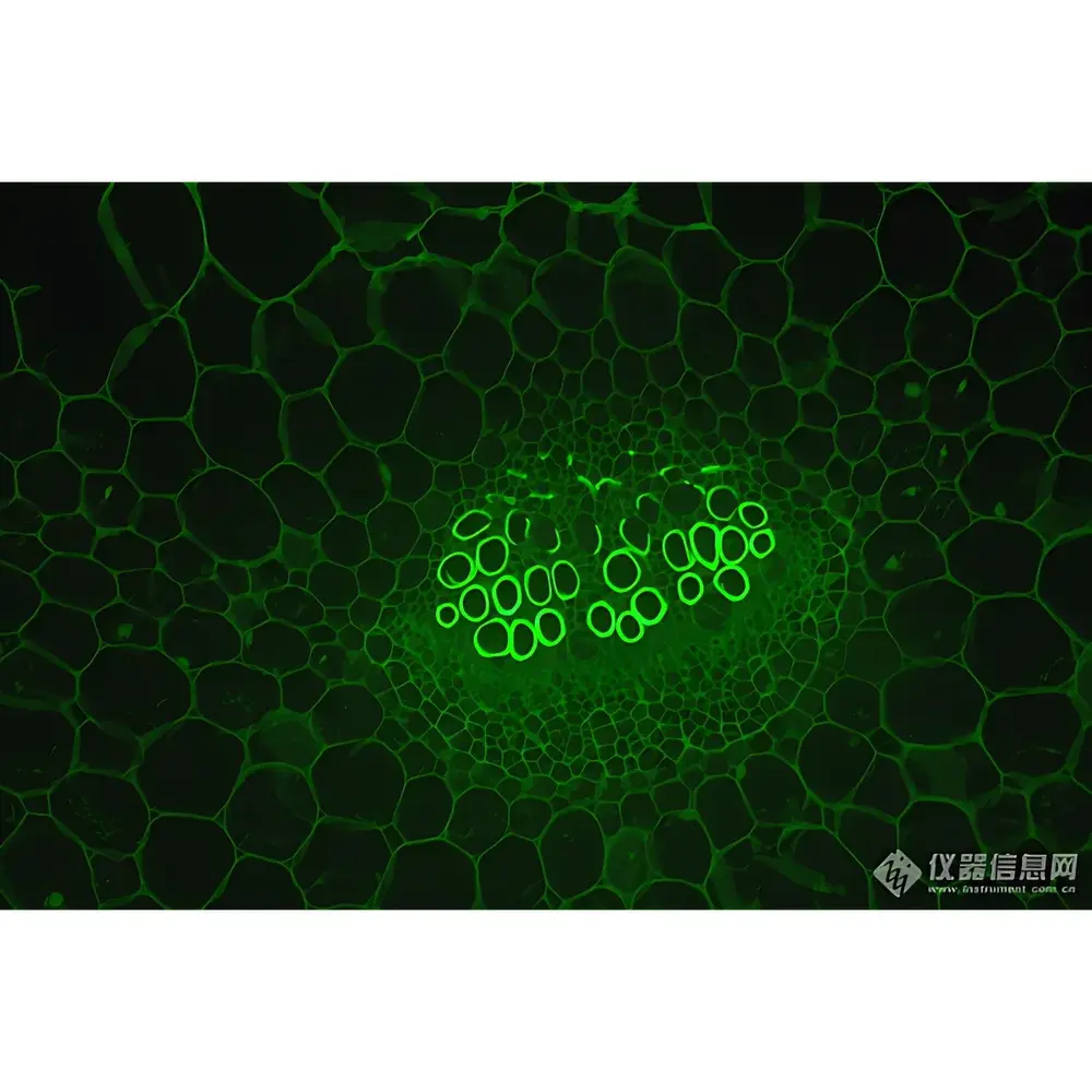
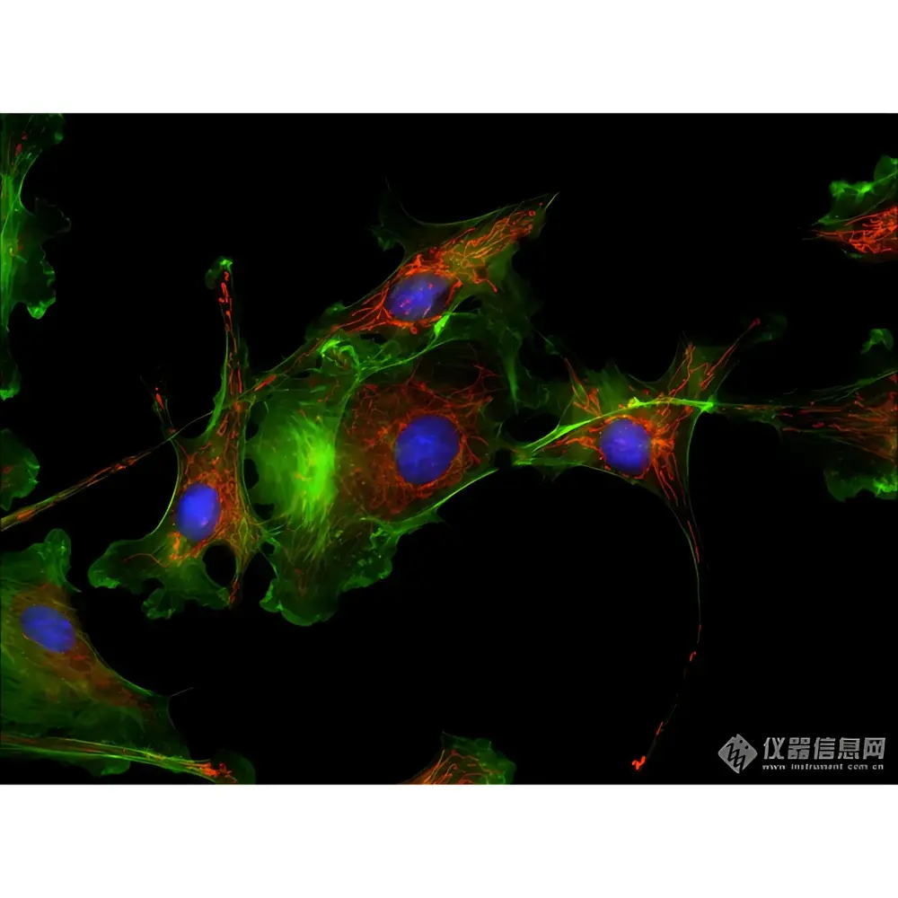
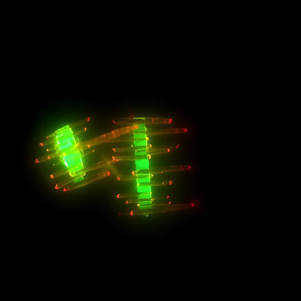
- Modular fluorescence turret accommodating up to four filter positions; B1 and G1 bandpass sets include excitation, dichroic, and emission filters matched to common fluorophores (e.g., DAPI, FITC, GFP).
- Integrated HDR (High Dynamic Range) image processing engine supporting real-time tone mapping, glare suppression, and local contrast enhancement—particularly beneficial for heterogeneous samples with high-intensity nuclei and low-signal cytoplasmic regions.
- Real-time extended depth-of-field (EDF) stacking and multi-field mosaic stitching capabilities via native firmware—enabling automated z-series acquisition and large-area tile scanning without external software dependency.
Sample Compatibility & Compliance
The LK-YG91 accommodates standard in vitro culture formats including Petri dishes (35–150 mm), multi-well plates (6–96-well), chambered coverglasses, and Terasaki plates—secured via optional stage adapters with precision alignment grooves. Its 72 mm achromatic condenser supports Köhler illumination optimization for both BF and PH modes, while the LED-based epi-illumination path delivers uniform excitation intensity across the field of view (FOV), minimizing phototoxicity during longitudinal assays. The system complies with IEC 61000-6-3 (EMC emissions) and IEC 61000-6-2 (immunity) standards. While not classified as a medical device (non-FDA 510(k)/CE-IVD certified), its optical performance meets ISO 10934-1 (microscope terminology) and ISO 9345-2 (objective lens labeling) requirements. For regulated environments, audit trails for image acquisition metadata (timestamp, exposure, gain, filter position) are exportable in CSV/TXT format to support internal SOP documentation and GLP traceability.
Software & Data Management
The microscope operates with LEI-TECH’s proprietary Imaging Suite v3.2—a Windows-based application supporting live streaming, time-lapse recording, region-of-interest (ROI) measurement (area, perimeter, intensity profiling), and calibrated pixel-to-micron conversion via stage calibration routines. All acquired images embed EXIF-style metadata (objective ID, magnification, illumination mode, exposure ms, gain dB), facilitating downstream analysis in ImageJ/Fiji, MATLAB, or commercial platforms like Imaris or Huygens. Raw TIFF output preserves 16-bit depth; JPEG exports include embedded ICC profiles for color consistency. The software enforces user-level access controls (admin/operator modes) and logs all parameter changes—aligning with basic FDA 21 CFR Part 11 principles for electronic records in non-clinical research settings. No cloud storage or remote telemetry is enabled by default; data remains fully local unless explicitly exported.
Applications
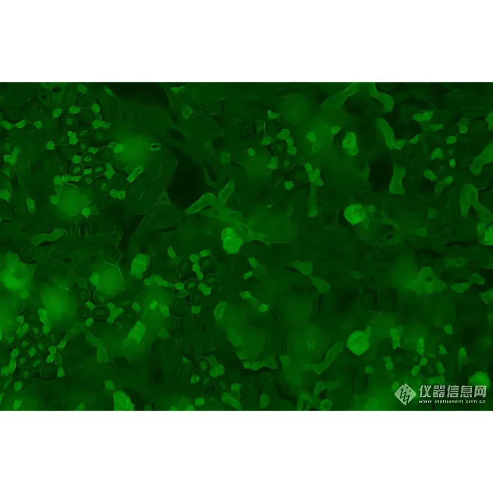
- Long-term live-cell imaging of fluorescently labeled organelles (mitochondria, lysosomes, ER) under physiological CO₂/humidity conditions using environmental chambers.
- Morphometric analysis of primary neuron cultures, including dendritic spine counting, axon length quantification, and growth cone dynamics.
- Validation of transfection efficiency and subcellular localization of GFP/RFP-tagged constructs in adherent cell models.
- Quality control of iPSC-derived cardiomyocyte monolayers via contractility assessment under phase contrast and calcium indicator (e.g., Fluo-4) fluorescence.
- Co-culture interaction studies (e.g., tumor–stroma, immune–cancer cell) using differential labeling and multi-channel overlay analysis.
- Teaching laboratory applications: comparative instruction on optical contrast mechanisms, fluorophore spectral properties, and resolution limits governed by Abbe’s criterion.
FAQ
Is the LK-YG91 compatible with third-party cameras?
Yes—it features a standardized C-mount interface (1× magnification relay) and supports USB 3.0/USB-C CMOS sensors up to 20 MP. Adapter rings for Canon EF, Nikon F, and Sony E-mount are available upon request.
Can the system be upgraded to support DIC or confocal modules?
The optical train includes预留 space for future DIC prism insertion; however, true confocal capability requires external laser scanning units and is not natively supported. Structured illumination (SIM) add-ons are under development.
What is the warranty coverage and service response time?
LEI-TECH provides a 24-month limited warranty covering parts and labor. Technical support is available 24/7 via encrypted email and remote desktop; on-site service dispatch is guaranteed within 72 business hours in Tier-1 cities across Asia, Europe, and North America.
Does the microscope meet ISO 19005-1 (PDF/A) archiving standards for image reports?
While the software does not generate PDF/A natively, exported TIFF + metadata CSV files comply with long-term archival best practices recommended by the NIH Data Sharing Policy and the FAIR principles (Findable, Accessible, Interoperable, Reusable).
Are fluorescence filter sets available for UV or far-red excitation?
B1 and G1 sets are standard; custom filter cubes for DAPI (UV), Cy5 (650 nm), or mCherry (587 nm) can be ordered separately with lead times of 4–6 weeks.
Related Products


