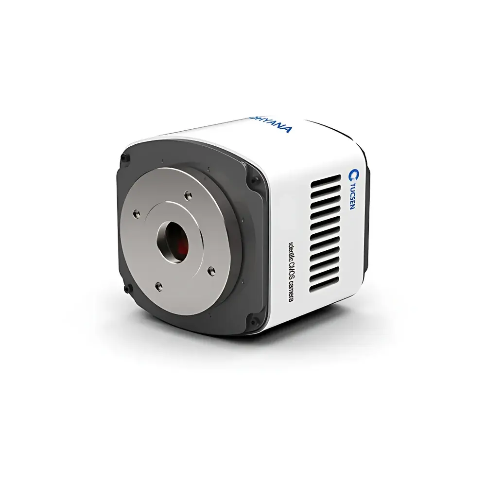
Tucsen Dhyana 400DC Scientific sCMOS Camera for Multimodal Microscopy (Fluorescence, Darkfield, DIC, Polarization)
| Brand | Tucsen |
|---|---|
| Origin | Fujian, China |
| Manufacturer Type | Original Equipment Manufacturer (OEM) |
| Product Category | Domestic |
| Model | Dhyana 400DC |
| Price | USD 7,000 (FOB) |
| Image Resolution | 4 MP (2048 × 2048) |
| Pixel Size | 6.5 µm × 6.5 µm |
| Sensor Format | 1.2″ |
| Readout Speed | 22 fps @ 8-bit, 16 fps @ 16-bit |
| Dynamic Range | 85 dB |
| Read Noise | 1.7 e⁻ (High Gain mode) |
| Full Well Capacity | 30,000 e⁻ |
| Cooling | Peltier-based thermoelectric cooling to −10 °C (ambient 25 °C) |
| Dark Current | 0.35 e⁻/pixel/s @ −10 °C (typ.) |
| Shutter | Rolling shutter |
| Exposure Range | 21 µs – 10 s |
| Bit Depth | 16-bit ADC |
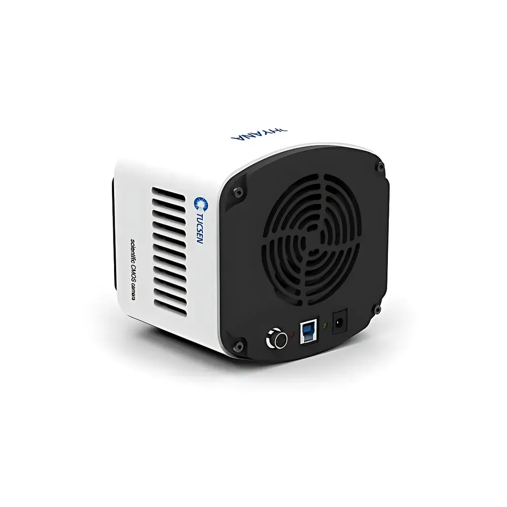
| Interface | USB 3.0 |
| Mount | C-mount |
| Dimensions | 120 × 119 × 121 mm |
| Power | 12 V / 8 A |
| Max. Power Consumption | 50 W |
Overview
The Tucsen Dhyana 400DC is a high-performance, color scientific complementary metal-oxide-semiconductor (sCMOS) camera engineered for multimodal optical microscopy applications—including epifluorescence, darkfield, differential interference contrast (DIC), and polarization imaging. Unlike conventional monochrome sensors requiring external filter wheels or sequential acquisition, the Dhyana 400DC integrates a back-illuminated, Bayer-patterned sCMOS sensor (G2020e) with on-chip color interpolation and a dedicated 16-bit image signal processor (ISP). This architecture enables simultaneous spectral fidelity and photon efficiency—critical for quantitative colocalization studies, live-cell dynamics under low-light conditions, and morphological analysis across multiple contrast modalities without hardware reconfiguration. Its 1.2″ optical format provides an expanded field of view compatible with standard microscope tube lenses and infinity-corrected objectives, reducing the need for image stitching while preserving spatial resolution and signal-to-noise ratio (SNR) across the full frame.
Key Features
- Ultra-low read noise: 1.7 electrons (high-gain mode), enabling reliable single-photon-level detection in weak-signal fluorescence and darkfield applications.
- True-color scientific imaging: Native Bayer array with human visual system (HVS)-weighted demosaicing ensures biologically faithful chromatic representation—essential for histopathology, pigment analysis, and multilabel immunofluorescence.
- 16-bit digitization with dual-gain architecture: Supports both high-dynamic-range (HDR) capture and high-speed acquisition (22 fps at 8-bit, 16 fps at 16-bit) without compromising linearity or quantitation accuracy.
- Vacuum-enhanced thermoelectric cooling: Stabilized at −10 °C (ambient 25 °C) via Peltier module with forced-air heat dissipation, suppressing dark current to ≤0.35 e⁻/pixel/s and maintaining thermal stability over extended acquisition sessions.
- USB 3.0 interface with zero-copy DMA transfer: Ensures deterministic latency and sustained bandwidth (>350 MB/s), fully compatible with real-time streaming, time-lapse synchronization, and hardware-triggered acquisition protocols.
- Comprehensive trigger support: Hardware-level triggering (standard, synchronous, global reset), programmable delay (0–10,000 s), and TTL-compatible output signals (exposure, global, readout) for integration into automated stages, light sources, or electrophysiology rigs.
Sample Compatibility & Compliance
The Dhyana 400DC is designed for use with standard upright and inverted research-grade microscopes equipped with C-mount adapters. Its 13.3 mm × 13.3 mm active area supports 40×–100× objective magnifications with minimal vignetting and optimal Nyquist sampling when paired with 0.21 µm–0.42 µm pixel-limited resolution optics. The camera complies with CE, FCC, and RoHS directives. Firmware and host software adhere to ISO/IEC 17025-relevant data integrity principles: raw image buffers are preserved unaltered in memory prior to ISP processing; metadata (exposure time, gain, temperature, timestamp) is embedded in TIFF/ND2 headers per MIAME and FAIR data guidelines. While not FDA 21 CFR Part 11-certified out-of-the-box, audit trails and user-access controls can be implemented via third-party LabArchives or Benchling integrations for GLP/GMP-aligned workflows.
Software & Data Management
The Dhyana 400DC ships with Mosaic—a cross-platform SDK supporting Windows and Linux—and offers native APIs for MATLAB, Python (via PyTucsen), LabVIEW, and Micro-Manager 2.0. All drivers expose low-level register access for custom pipeline development. The 16-bit ISP engine performs real-time flat-field correction, defect pixel replacement, and gamma-linear scaling—configurable per acquisition session without post-processing overhead. Export formats include uncompressed TIFF (with embedded EXIF/XMP), ND2 (Nikon), and HDF5 for large-scale volumetric or time-series datasets. Metadata conforms to OME-TIFF schema, ensuring compatibility with QuPath, Fiji/ImageJ, and commercial digital pathology platforms. Batch acquisition scripts support scheduled exposure ramping, Z-stack tiling, and multi-channel time-lapse with hardware synchronization.
Applications
- Quantitative fluorescence co-localization in fixed and live specimens (e.g., GFP/mCherry dual labeling, FRET acceptor photobleaching).
- Label-free morphological phenotyping using polarization contrast for birefringent structures (collagen, starch granules, liquid crystals).
- Dynamic darkfield imaging of nanoparticle tracking, bacterial motility, or plasmonic scattering events.
- DIC-based subcellular organelle segmentation in unstained primary neurons or epithelial monolayers.
- Materials science inspection: grain boundary mapping in metallurgical samples, thin-film interference analysis, and stress-induced birefringence in polymer composites.
FAQ
Is the Dhyana 400DC suitable for super-resolution microscopy techniques such as SIM or STORM?
Yes—the camera’s low read noise, high quantum efficiency (>75% peak), and precise timing control make it compatible with structured illumination and single-molecule localization microscopy when used with appropriate excitation modulation and stage synchronization.
Does the camera support global shutter operation?
No—it employs a rolling shutter; however, its fast readout (21 µs minimum exposure) and hardware-triggered global reset mode minimize motion artifacts in most biological and materials imaging scenarios.
Can I perform real-time background subtraction or flat-field correction during acquisition?
Yes—Mosaic and the SDK provide programmable ROI-based background estimation and multiplicative flat-field correction applied in the FPGA before 16-bit digitization.
What is the maximum sustainable frame rate at full resolution and 16-bit depth?
16 frames per second, with full-frame readout, no binning, and lossless compression disabled—verified under sustained USB 3.0 bandwidth and host memory allocation ≥4 GB.
Is there a Linux driver available for headless server deployment?
Yes—Tucsen provides a kernel-module-free userspace driver (libtucsen) with Python bindings and CLI tools for remote acquisition, ideal for HPC cluster-integrated imaging pipelines.


