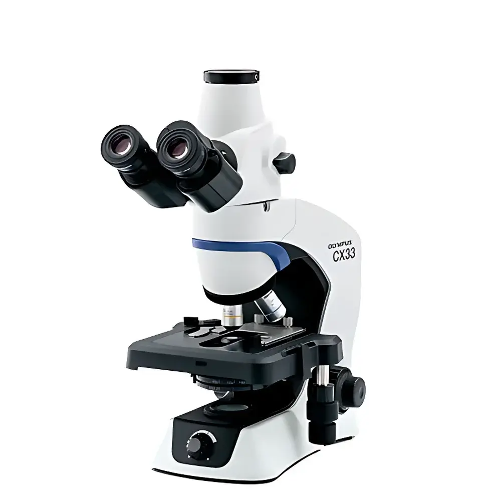
Olympus CX33 Biological Microscope
| Brand | Olympus |
|---|---|
| Origin | Guangdong, China (Distributed by Authorized Distributor) |
| Microscope Type | Upright Biological Microscope |
| Optical System | UIS2 Infinity-Corrected Optics |
| Illumination | Integrated LED Köhler Illumination (2.4 W, Daylight Color Temperature ~5500 K, 60,000 hr Lifetime) |
| Objective Turret | Fixed 4-Position Nosepiece |
| Stage | Mechanical Stage (211 mm × 154 mm), X–Y Travel: 76 mm × 5 mm, Single/Optional Dual Slide Holder with Scale and XY Stop |
| Viewing Tube | 30° Inclined Trinocular Tube (Anti-Fungal Coating), Beam Splitter Ratio: 100:0 or 0:100 (Eyepiece:Camera), Interpupillary Adjustment: 48–75 mm, Eye Point Adjustment: 375.0–427.9 mm |
| Objectives | Plan Achromatic (Anti-Fungal), 4×, 10×, 20×, 40×, 100× Oil |
| Eyepieces | Widefield 10× (FN 20 mm), Optional 15× (FN 16 mm) |
| Optional Accessories | Darkfield Condenser, C-Mount CCD Adapter, Ergonomic Eyepiece Extender, Dual-Head Teaching Attachment |
| Compliance | Designed to meet ISO 10993-5 biocompatibility requirements for optical components |
Overview
The Olympus CX33 is a high-reliability upright biological microscope engineered for routine histology, cytology, microbiology, and academic teaching applications. Built upon the proven UIS2 (Universal Infinity System) optical platform, it delivers consistent, flat-field image quality across its full magnification range—from 40× to 1000×—with minimal chromatic and spherical aberration. Its infinite-corrected optical path enables seamless integration of auxiliary modules (e.g., darkfield condensers, fluorescence filter cubes via optional adapters) without compromising resolution or contrast. The microscope employs Köhler illumination architecture with a precisely aligned, built-in LED light source that maintains stable color temperature (~5500 K) across all intensity settings—eliminating white balance recalibration during brightness adjustment and ensuring faithful representation of natural specimen coloration under standardized daylight-equivalent conditions.
Key Features
- Ergonomic mechanical stage with low-profile design: 211 mm × 154 mm footprint and 76 mm × 5 mm travel range allows intuitive one-handed specimen positioning while maintaining forearm support on the stage surface.
- Fixed 4-position objective nosepiece with smooth, low-torque rotation—optimized for rapid, fatigue-free magnification switching during extended observation sessions.
- LED illumination system rated for 60,000 hours: Provides stable luminance output over lifetime, eliminating filament degradation and thermal drift common in halogen sources—critical for quantitative brightfield imaging reproducibility.
- Plan achromatic objectives with anti-fungal coating: Delivers uniform sharpness across the entire field of view (FN 20 mm), essential for accurate morphometric analysis and slide screening efficiency.
- Trinocular viewing tube with 100:0 / 0:100 beam splitter: Enables simultaneous visual inspection and digital capture via C-mount camera interface without optical path compromise.
- Dual-slide holder option and calibrated stage controls: Supports high-throughput workflow in clinical labs and educational settings where multi-specimen comparison is routine.
Sample Compatibility & Compliance
The CX33 accommodates standard glass microscope slides (76 mm × 26 mm), blood smear preparations, tissue sections (paraffin-embedded or frozen), live unstained cells (phase contrast compatibility via optional condenser), and microbiological smears. Its stage geometry and specimen clamping system are optimized for both single and dual-slide configurations—including hemocytometer use. All optical components comply with ISO 10993-5 standards for cytotoxicity testing of medical device materials, confirming biocompatibility of lens coatings and housing surfaces. While the instrument itself does not carry CE IVD or FDA 510(k) clearance as a standalone diagnostic device, its optical performance and mechanical stability align with ASTM E2877-22 guidelines for routine brightfield microscopy validation in QC/QA environments. When paired with FDA 21 CFR Part 11–compliant imaging software and audit-trail-enabled acquisition hardware, the system supports GLP and GMP-aligned documentation protocols.
Software & Data Management
The CX33 does not include embedded firmware or proprietary acquisition software; instead, it interfaces seamlessly with third-party digital imaging platforms via standard C-mount (1× magnification factor) and USB-powered camera adapters. Compatible solutions include Olympus cellSens Entry (for basic annotation and measurement), Media Cybernetics Image-Pro Premier (for quantitative morphometry), and open-source alternatives such as Fiji/ImageJ with appropriate calibration routines. Metadata capture—including objective magnification, illumination intensity, and timestamp—is fully supported when using cameras with EXIF-compliant drivers. For regulated environments, integration with LIMS or ELN systems is achievable through DICOM-SR or CSV export pipelines, provided the host imaging software implements electronic signature and audit trail functionality per 21 CFR Part 11 requirements.
Applications
- Routine histopathology screening in hospital laboratories and reference centers
- Microbiology identification and Gram staining evaluation
- Hematology analysis including peripheral blood smear review and reticulocyte counts
- Academic instruction in undergraduate biology and medical training programs
- Quality control of cell culture morphology in biomanufacturing support labs
- Plant anatomy and protist observation in environmental research
FAQ
Is the CX33 suitable for oil immersion microscopy?
Yes—the included 100× plan achromatic objective is designed for oil immersion use and features spring-loaded retraction to prevent damage during focusing.
Can the CX33 be upgraded for phase contrast or fluorescence?
Phase contrast requires an optional phase contrast condenser and matching phase objectives; fluorescence capability is not natively supported but may be added via third-party LED epi-illuminators and filter cubes with appropriate adapter rings.
What is the maximum usable magnification with the standard eyepieces?
With 10× widefield eyepieces and the 100× oil objective, total magnification reaches 1000×—within the practical resolution limit of visible-light brightfield microscopy (~0.2 µm at 550 nm wavelength).
Does the LED illumination support intensity modulation for phototoxicity-sensitive live samples?
Yes—the continuous dimming control allows precise irradiance reduction down to <1% nominal output, enabling low-light observation of unstained or fluorescently labeled living specimens.
Is service and calibration support available outside Japan?
Authorized Olympus service partners operate globally; calibration certificates traceable to NIST standards are issued upon request for ISO/IEC 17025–accredited facilities.


